
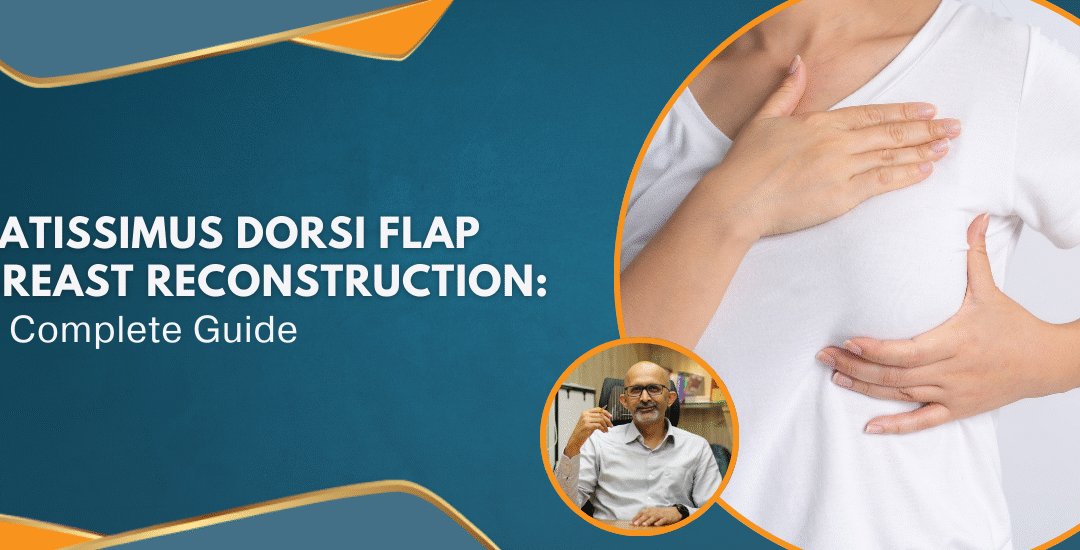
Latissimus Dorsi Flap Breast Reconstruction: A Complete Guide
What is Latissimus Dorsi Flap Breast Reconstruction?

Think you can use your own skin and muscle to restore the natural shape of your breast. That is precisely what latissimus dorsi flap for breast reconstruction achieves.
The LD flap reconstruction employs the latissimus dorsi muscle, found in your upper back, to form a new breast mound. Part of this muscle, along with fat and skin, is brought to the chest with its blood supply intact. The procedure can be done with or without an implant, depending on the volume required for reconstruction.
How is the Procedure Performed?

Step 1: A portion of the latissimus dorsi muscle, skin, and fat is separated while leaving its blood supply intact.
Step 2: The tissue is tunneled from under the arm to the chest.
Step 3: The tissue is molded by the surgeon to create a new breast.
Step 4: If needed, an implant is inserted to create desired volume.
This technique is particularly beneficial for patients lacking sufficient abdominal fat for other flap-based reconstructions such as the DIEP or TRAM flaps.
Read on to know if LD flap reconstruction is a preferred option nowadays.
Is the LD Flap Suitable for Breast Reconstruction?
Now, let’s understand the advantages and disadvantages of the LD flap.
Latissimus Dorsi Flap Reconstruction: Pros and Cons
Benefits
- High success rate – The LD flap has a very low failure rate, so it’s a good choice.
- Suitable for thin patients – If you don’t have sufficient abdominal fat for a DIEP or TRAM flap, this method can be a great alternative.
- Can be used with implants – If more volume is desired, the flap can be done with an implant.
- Few donor-site complications – The back heals well after the muscle is transferred.
Drawbacks
- Loss of muscle function – Although not a significant problem for most, athletes who use upper body strength may experience some weakness.
- Feels firmer than natural breast tissue – The muscle-based reconstruction may not feel as natural as fat-based approaches.
- Potential scarring – There will be a scar on the back, although it is often concealed by clothes.
Are you worried about the risks?
Possible Risks Involved with LD Flap Reconstruction
Although LD flap reconstruction is considered safe, as with any surgery, it does involve some risks, which include:
- Fluid buildup (seroma) – Fluid may accumulate in the back, necessitating drainage.
- Infection – As with any surgery, there’s a small risk of infection.
- Weakness in upper body movement – Some patients may experience reduced strength in the shoulder and back.
- Scar formation – Both on the back (donor site) and chest (reconstructed breast).
Following your surgeon’s post-operative care instructions can help minimize these risks and promote a smoother recovery.

 Natural products have been used in medicine for centuries. From traditional herbal remedies to plant-based extracts, many believe that nature holds the key to curing diseases, including cancer. But how do these natural substances interact with cancer treatments?
 Myth 1: Natural products have no side effects
 One of the biggest risks with tree bark medicine for cancer is the lack of dosage control. Unlike pharmaceutical drugs, which are carefully measured, natural products vary in potency. Consuming too much can cause poisoning, while too little may have no effect.
 Cancer is a complex disease, and the search for a cure is ongoing. Understandably, patients seek hope in natural cancer treatment, especially when modern medicine feels overwhelming. However, unverified remedies should not replace scientifically proven treatments. The use of tree bark medicine for cancer or other plant-based therapies may seem appealing, but they carry risks that one cannot ignore.
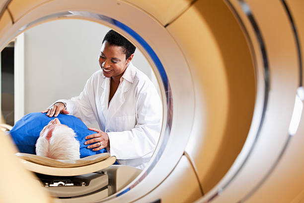 Positron Emission Tomography (PET) scan, often combined with a Computed Tomography (CT) scan, is an advanced imaging technique that detects abnormal metabolic activity in the organs of the body. Commonly it involves injecting a small amount of radioactive glucose (FDG), which cancer cells absorb at a higher rate than normal cells, making them visible on the scan. There are other radioactive traces that are specific for some cancers like PSMA for prostate cancer and Dota for Neuroendocrine tumors.
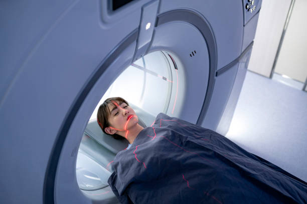 1. It’s a Painless and Non-Invasive Procedure
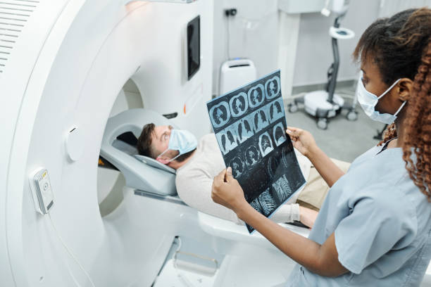 1. Early and Accurate Detection
