
This website is being reviewed and updated in line with the National Medical Commission (NMC) guidelines on ethical advertising and public communication by registered medical practitioners.
support@macsclinic.com
+91 9482202240
First Week After Cancer Diagnosis?

First Week After Cancer Diagnosis?

The first week isn’t for treatment. It’s for getting your footing. The priorities are clear: confirm the diagnosis is solid, gather every report and scan in one place, and get in front of a specialist who treats your specific cancer. Most cancers allow that window, and using it well shapes every decision that follows.

According to Prof. Dr. Sandeep Nayak, Surgical Oncologist in India, “The first week should be spent confirming the diagnosis and understanding the options, not panicking into a treatment that hasn’t been properly planned, because a rushed start rarely improves anything.”

Feeling lost in the first few days?

What Should You Actually Do in the First Few Days?

Nobody hands you a checklist with the diagnosis. So here’s where the energy should go first.

  • Gather records: Pull your biopsy, imaging and blood reports into one file. Every specialist you see will want them to weigh in properly.
  • Find the right specialist: A doctor who treats your exact cancer at volume reads the case differently. That gap shows up in the plan you’re offered.
  • Hold off on panic: It feels like an emergency, yet rushing into surgery within days rarely helps and can lock you into the wrong path.
  • Bring someone along: A second set of ears catches what you’ll miss. Nobody absorbs much in the appointment right after hearing the news.

None of this slows treatment down. It’s groundwork, and patients heading toward robotic cancer surgery start from a far stronger position when the first week is handled well.

When Should You Get a Second Opinion?

Early, ideally within this first week, and for solid reasons. Here’s why it’s worth doing now.

  • Confirms the diagnosis: Another specialist checking the pathology and staging can catch the rare error before it shapes your entire treatment.
  • Opens up options: A fresh set of eyes sometimes surfaces approaches the first consultation never raised, which matters when treatments carry lasting consequences.
  • Before treatment starts: Once chemo or surgery is underway, your choices narrow fast, so the time to look around is now, not later.
  • Settles the fear: Even when the second view agrees, knowing the plan has been tested takes a lot of weight off those early days.

So it isn’t a sign of distrust. The same thinking behind a second opinion at any stage applies here, get the diagnosis right before committing to anything.

Why Choose Dr. Sandeep Nayak for Your Cancer Diagnosis?

Dr. Sandeep Nayak brings 24 years of surgical oncology experience, DNB qualifications in Surgical Oncology and General Surgery and a fellowship in Laparoscopic and Robotic Onco-Surgery to the care of newly diagnosed patients across all cancer types. He sees patients personally in those first overwhelming days and explains plainly what the diagnosis means and what the realistic options are.

That early clarity is what turns panic into a plan. Every case at MACS Clinic goes through a full tumour board, where imaging, pathology and oncology weigh in together before anything is confirmed. Call +91 8104310753 to book your consultation.

Frequently Asked Questions

What should I do first after a cancer diagnosis?

Confirm the diagnosis, gather all reports and see a specialist who treats your specific cancer.

Should I get a second opinion in the first week?

Yes, a second opinion early on confirms the plan before any treatment begins.

Do I need to start treatment immediately?

Rarely within days, most cancers allow time to plan properly without losing ground.

What records should I collect?

Biopsy, imaging, blood tests and any prior medical history relevant to the diagnosis.

References:

  1. National Cancer Institute — Newly Diagnosed With Cancer. https://www.cancer.gov/
  2. World Health Organisation — Cancer. https://www.who.int/news-room/fact-sheets/detail/cancer
What Does a Suspicious Biopsy Mean?

What Does a Suspicious Biopsy Mean?

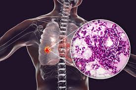
A suspicious biopsy report sits in the grey zone between clearly benign and confirmed cancer. It means the pathologist saw cells that look abnormal, enough to raise a flag, but not enough to call it cancer with certainty. So it isn’t a diagnosis. It’s a signal that more is needed, usually further testing or a repeat sample, before anyone can say for sure either way.

According to Prof. Dr. Sandeep Nayak, Surgical Oncologist in India, “A suspicious report isn’t bad news or good news yet, it’s a call to investigate properly, and I’d far rather act on a careful follow-up than treat or dismiss something on a guess.”

Got a report you can’t quite make sense of?

Why Does a Biopsy Come Back Suspicious?

A few different things land a report in that uncertain middle. Here’s what’s usually behind it.

  • Too few cells: When the sample is sparse or poorly preserved, the pathologist can see something’s off but doesn’t have enough to commit to a verdict.
  • Borderline appearance: Some cells sit right between normal and malignant, showing early changes that could go either way without more information.
  • Overlapping features: Certain benign conditions mimic cancer closely under the microscope, and telling them apart often needs more than a first look.
  • Sampling spot: If the needle caught the edge rather than the core of a lump, the most telling cells may simply not be in the sample.

So “suspicious” often says more about the sample than the disease. Anyone heading toward robotic cancer surgery needs that uncertainty resolved first, not carried into the operating room.

What Happens After a Suspicious Result?

The report isn’t the finish line, it’s a prompt for the next step. This is what usually follows.

  • Special staining: Pathologists apply molecular tests that tag specific proteins, turning a borderline read into a much clearer answer about the cell type.
  • Repeat biopsy: Sometimes the simplest fix is a fresh sample, ideally a core biopsy that pulls more tissue than the first attempt managed.
  • Expert review: A second pathologist, often a specialist in that cancer type, can settle a difficult case that one reading left open.
  • Clinical correlation: The result gets weighed against your scans and symptoms, because a report never stands entirely on its own.

So uncertainty is temporary, not permanent. Much like what an IHC test adds to the picture, the next test is what turns “suspicious” into something you can actually act on.

Why Choose Dr. Sandeep Nayak for Your Cancer Diagnosis?

Dr. Sandeep Nayak has spent 24 years in surgical oncology, with DNB qualifications in Surgical Oncology and General Surgery and a fellowship in Laparoscopic and Robotic Onco-Surgery. He’s spent much of that time in exactly these diagnostic grey zones, where a report and a clinical picture don’t quite line up and someone has to decide the next move.

And that judgment is what keeps a suspicious report from becoming either overtreatment or a missed cancer. Every case at MACS Clinic runs through a full tumour board, where pathology, imaging and oncology weigh in together before anything is confirmed. Call +91 8104310753 to book your consultation.

Frequently Asked Questions

Does a suspicious biopsy mean I have cancer?

Not always, it means cancer is possible but not yet confirmed.

What happens after a suspicious biopsy?

Usually further testing like IHC or a repeat biopsy to reach certainty.

How long until I get a clear answer?

Often within a week or two, depending on the additional tests needed.

Should I get a second opinion?

Yes, a suspicious report is a sensible point to seek expert review.

References:

      1. National Cancer Institute — Pathology Reports. https://www.cancer.gov/
      2. World Health Organisation — Cancer. https://www.who.int/news-room/fact-sheets/detail/cancer
How Accurate Is FNAC for Cancer?

How Accurate Is FNAC for Cancer?

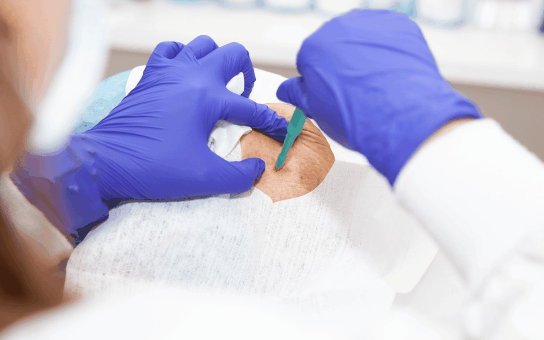
FNAC is surprisingly accurate for what it is, a thin needle drawing a few cells from a lump. In experienced hands it gets the answer right well over ninety percent of the time for many cancers, especially in the thyroid, breast and lymph nodes. It isn’t flawless though, and when the result is unclear or the sample’s too thin, a core biopsy usually takes over to settle it.

According to Prof. Dr. Sandeep Nayak, Surgical Oncologist in India, “I trust FNAC for a thyroid or lymph node lump, but I never let a single negative result overrule a lump that looks and feels like cancer, that’s when I push straight to a core biopsy.”

Unsure if your FNAC result is the full answer?

What Makes FNAC Accurate or Inaccurate?

Accuracy isn’t fixed. It swings a lot depending on a few things going right.

  • Who does it: A skilled hand placing the needle in the right spot changes everything, since a sample drawn from the edge of a lump can miss the cancer entirely.
  • Cell quality: The pathologist needs enough clear cells to judge, and a sparse or bloody sample is where false negatives tend to creep in.
  • Cancer type: Some tumours shed cells that read easily, while others, like certain follicular thyroid cancers, simply can’t be confirmed by cells alone.
  • The lump itself: Tiny, deep or hard-to-reach lumps are harder to sample cleanly, which is exactly when accuracy starts to dip.

So a “negative” FNAC isn’t always the end of it. Anyone weighing up robotic cancer surgery wants that diagnosis nailed down first, not left on a borderline result.

When Do You Need More Than an FNAC?

Sometimes cells alone don’t cut it. Here’s when your doctor reaches for the next test.

  • Unclear result: If the FNAC comes back inconclusive or suspicious rather than definite, a core biopsy gives the tissue needed to be sure.
  • Architecture matters: FNAC shows cells, not how they’re arranged, and for some cancers that arrangement is what confirms the type.
  • Planning treatment: Detailed tests like hormone receptors or molecular markers often need a tissue sample, which FNAC can’t always provide.
  • Result and clinic clash: When a clean FNAC sits next to a worrying lump or scan, that mismatch is a clear signal to dig deeper.

So FNAC and tissue testing aren’t rivals, they work in sequence. Much like the choice explained in our core biopsy comparison, it’s about matching the test to what the case actually needs.

Why Choose Dr. Sandeep Nayak for Your Cancer Diagnosis?

Dr. Sandeep Nayak has spent 24 years in surgical oncology, with DNB qualifications in Surgical Oncology and General Surgery and a fellowship in Laparoscopic and Robotic Onco-Surgery. He reads an FNAC report the way a surgeon has to, never in isolation, always against the lump, the scans and the bigger clinical picture.

And that’s what stops a borderline result from becoming a wrong turn. Every case at MACS Clinic runs through a full tumour board, where pathology, imaging and oncology weigh in together before anything is confirmed. Call +91 8104310753 to book your consultation.

Frequently Asked Questions

How accurate is FNAC for cancer?

FNAC is highly accurate for many cancers, often above ninety percent in skilled hands.

Can FNAC miss cancer?

Yes, a false negative can happen if the needle misses the cancerous cells.

Is FNAC enough to confirm cancer?

Often yes, but unclear results may need a core biopsy for confirmation.

Does FNAC hurt?

It causes only mild discomfort, similar to a routine blood test.

References:

  1. National Cancer Institute — Fine Needle Aspiration. https://www.cancer.gov/
  2. World Health Organisation — Cancer. https://www.who.int/news-room/fact-sheets/detail/cancer
    Why Does a Doctor Want a PET Scan After a CT Scan?

    Why Does a Doctor Want a PET Scan After a CT Scan?

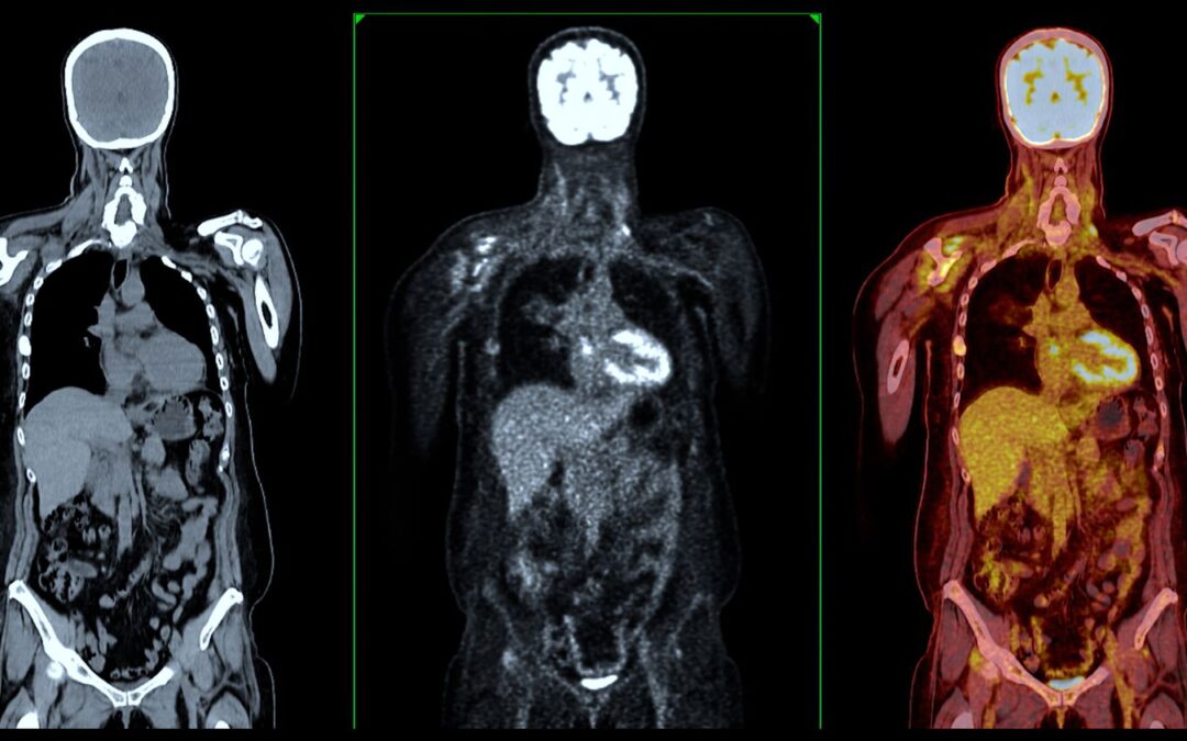
    A doctor orders a PET scan after a CT because the two tests answer different questions. A CT shows the size, shape and location of a mass, while a PET reveals how metabolically active that tissue actually is. Cancer cells burn glucose fast, so they light up on a PET in a way a CT simply can’t show. Together they confirm whether the disease is active and how far it has spread.

    According to Prof. Dr. Sandeep Nayak, Surgical Oncologist in India, “A CT tells me what a lump looks like, but a PET tells me whether it’s behaving like cancer, and that difference often decides the whole treatment plan.”

    Unsure why one scan isn’t enough? Book An Appointment

    What Does a PET Scan Show That a CT Scan Cannot?

    A CT maps anatomy. A PET maps activity. That gap is the entire reason both get used.

    • Cell activity: PET picks up the high glucose uptake of cancer cells, so it flags disease that’s biologically active rather than just a shadow on an image.
    • Hidden spread: Small deposits in lymph nodes or distant organs often stay invisible on CT, and PET catches many of them before they grow large enough to see.
    • Scar vs cancer: After treatment, a CT can show a lingering mass that’s only scar tissue, and PET tells whether it’s dead or still alive.
    • Whole body: One PET scan surveys the entire body at once, which matters when the worry is spread rather than a single known site.

    So the two aren’t rivals. A patient working through their cancer staging gets a far more complete picture when structure and activity are read side by side.

    When Is a PET Scan Actually Necessary After a CT?

    Not every case needs one. But in specific situations a PET changes the decision entirely.

    • Staging: When a cancer is confirmed, PET helps pin the true stage by checking whether it has quietly travelled beyond the primary site.
    • Unclear findings: If a CT shows something borderline that could go either way, PET often settles whether it’s worth a biopsy or surgery.
    • Treatment response: Midway through chemo or radiation, PET shows whether the tumour is genuinely shrinking in activity, not just in size.
    • Suspected recurrence: When markers rise but a CT looks clean, PET can locate disease that’s returned before anything else picks it up.

    So the timing isn’t random. Much like getting a second opinion, the extra scan is about confirming the picture before committing to a plan.

    Why Choose Dr. Sandeep Nayak for Your Cancer Diagnosis?

    Dr. Sandeep Nayak holds a DNB in Surgical Oncology and General Surgery, with 24 years in the field and a practice built entirely around cancer. He reads imaging the way a surgeon has to, looking not just at what a scan shows but at what it means for whether, when and how to operate.

    For a patient, that’s the difference between a scan report and a plan. Every case at MACS Clinic goes through a full tumour board, where imaging, pathology and oncology are weighed together before anything is confirmed. Reach the team at 📞 +91 9482202240.

    Frequently Asked Questions

    Is a PET scan always needed after a CT?

    No, it’s ordered only when staging, unclear findings or suspected spread make it useful.

    Does a PET scan confirm cancer on its own?

    No, it shows activity, but a biopsy is still needed for a definite diagnosis.

    Is a PET scan safe?

    Yes, it uses a low dose of short-lived radioactive tracer that clears quickly.

    How long does a PET scan take?

    Usually around two to three hours, including the tracer uptake waiting period.

    References:

    1. National Cancer Institute — PET Scans. https://www.cancer.gov/
    2. World Health Organisation — Cancer. https://www.who.int/news-room/fact-sheets/detail/cancer

    Why Robotic Surgery is the Future of Cancer Treatment?

    Why Robotic Surgery is the Future of Cancer Treatment?

    Robotic surgery is becoming the future of cancer treatment because it gives the surgeon precision, magnified vision and reach that open and laparoscopic methods can’t match consistently. Tumours come out through small incisions, with nerves and healthy tissue left intact, so pain drops and recovery speeds up. And for many solid tumours, the outcome data already leans this way.

    According to Prof. Dr. Sandeep Nayak, Surgical Oncologist in India, “Robotic surgery lets us operate in spaces the human hand can’t reach cleanly, and that control is exactly what changes a difficult cancer case into a safe one.”

    Wondering if robotic surgery is right for your cancer?

    What Makes Robotic Surgery Better Than Traditional Cancer Surgery?

    The edge isn’t the machine itself. It’s what the surgeon can suddenly do.

    • 3D vision: The surgeon works from a magnified high-definition 3D view, not the flat 2D image laparoscopy offers, so depth and tissue planes stay clear right through the operation.
    • Motion control: Instrument movement scales down to a fraction of the hand’s motion, which settles natural tremor and makes fine dissection possible in tight, awkward spots.
    • Nerve sparing: Veins and nerves show up enlarged and far easier to spot, and that’s decisive in rectal, prostate and gynaecologic surgery where preserving them matters.
    • Smaller wounds: A few sub-centimetre cuts replace one long incision, so blood loss falls and patients are back on their feet much sooner.

    And those gains stack up. Anyone weighing robotic cancer surgery should look at the surgeon’s experience just as hard as the technology.

    Which Cancers Benefit Most From Robotic Surgery?

    Not every tumour needs it. But several are a genuinely strong fit.

    • Prostate cancer: The gland sits deep in the pelvis, surrounded by nerves that control continence and potency, and robotic access protects them more reliably than open surgery does.
    • Colorectal cancer: These cases are technically tough, and nerve-sparing robotic technique helps preserve bladder, bowel and sexual function wherever the disease allows.
    • Gynaecologic cancers: Ovarian, cervical and uterine tumours sit in cramped pelvic anatomy, and the wristed instruments reach angles rigid laparoscopic tools simply can’t.
    • Head and neck cancer: Transoral robotic surgery reaches throat tumours through the mouth, so there’s no cut across the face or jaw and recovery is gentler.

    So the real question isn’t whether the technology looks impressive. It’s whether your specific cancer is one where it actually changes the result. Our blog on robotic cancer surgery costs breaks down when it’s worth it.

    Why Choose Dr. Sandeep Nayak for Robotic Cancer Surgery?

    Dr. Sandeep Nayak trained overseas specifically to master laparoscopic and robotic onco-surgery, holds a DNB in Surgical Oncology and General Surgery, and has performed hundreds of robotic cancer procedures across 24 years in the field. He’s also the originator of three published techniques, RABIT, MIND and RIA-MIND, which surgeons now travel to learn.

    For a patient, what that adds up to is simple. You aren’t getting someone who tries robotics now and then. You’re getting one of the most experienced robotic cancer surgeons in the country, with every case run past a full tumour board before anything gets confirmed. Reach the team at 📞 +91 9482202240.

    Frequently Asked Questions

    Does a robot perform the surgery on its own?

    No, the surgeon controls every movement from a console, the robot cannot act independently.

    Is robotic cancer surgery safe?

    Yes, in trained hands it offers less blood loss, lower infection risk and reliable outcomes.

    How long is recovery after robotic surgery?

    Most patients recover faster than open surgery, with shorter hospital stays and quicker return to routine.

    Can robotic surgery treat advanced cancers?

    Yes, it treats many advanced solid tumours, though suitability depends on stage and location.

    References

    1. National Cancer Institute — Robotic Surgery. https://www.cancer.gov/
    2. World Health Organisation — Cancer. https://www.who.int/news-room/fact-sheets/detail/cancer

    Call Now Button