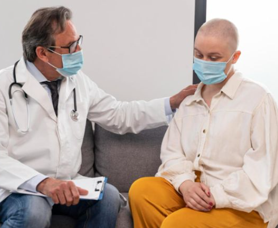
Immune Recovery After Cancer Surgery
Weeks. Sometimes months. And almost always longer than patients expect when they walk out of hospital thinking the hard part is behind them. The wound closes in weeks. Everyone can see that healing happening. But the immune system works somewhere nobody can see and it takes its own time on its own timeline regardless of how well the scar looks from the outside.
According to Dr. Sandeep Nayak, surgical oncologist in India, “The wound healing and the immune recovery are two completely different processes and conflating them is why so many patients are genuinely blindsided by how vulnerable they feel months after surgery.”
What Is Surgery Actually Doing to Your Immune System?
Most people think the immune hit from surgery is about the wound. It isn’t. It’s bigger than that and it runs deeper than that.
- Your Body Interprets Surgery as Catastrophic Trauma and Pulls Every Available Immune Resource Toward Survival: Natural killer cells. Lymphocytes. The entire surveillance system that normally patrols your body looking for abnormal cells. All of it gets redirected toward wound healing and tissue repair leaving you genuinely more vulnerable to infection and less capable of immune surveillance for weeks.
- The Anaesthetic Itself Suppresses Immune Function Independently of Everything the Surgery Is Doing: Anaesthetic agents dampen natural killer cell activity and neutrophil function for days after you wake up and that effect stacks on top of the surgical trauma happening simultaneously rather than replacing it.
- Lymph Node Removal Doesn’t Just Temporarily Reduce Immune Capacity. It Permanently Changes the Architecture: When lymph nodes come out as part of cancer treatment the regional surveillance network loses capacity that doesn’t grow back and this isn’t a temporary dip. It’s a permanent restructuring of how that part of your immune system works going forward.
- The Nutritional Crash After Major Surgery Starves the Immune System at the Exact Moment It Needs Feeding: Protein stores drop. Zinc drops. Vitamin D drops. Vitamin C drops. And an immune system trying to rebuild itself without the raw materials it needs is like a construction crew trying to build a house with no bricks delivered. It takes longer. Much longer.
In cases of cancers where a high degree of accuracy in tumour removal is demanded in anatomically complex regions, innovative robotic surgery technologies are becoming a popular method of enhancing the accuracy of surgery and recovery in patients.
What Makes Recovery Faster for Some People and Slower for Others?
These aren’t random differences. They’re driven by specific variables that you can understand and in many cases actively influence.
- A Robotic Minimally Invasive Operation Creates a Shallower Immune Suppression Than Open Surgery From Day One: Less surgical trauma means less immune disruption means a shorter recovery window and this is one of the most clinically meaningful advantages of minimally invasive approaches that rarely gets explained to patients before they choose their surgeon.
- Chemotherapy Starting Soon After Surgery Doesn’t Give the Immune System Time to Find Its Feet Again: The immune system that was slowly climbing back gets knocked down again by the first chemotherapy cycle and genuine recovery can only begin after the absolute last cycle of treatment is completely done.
- Age Determines How Much Reserve the Immune System Has to Draw on During the Rebuilding Process: A 35 year old and a 68 year old having identical operations will not have identical immune recovery timelines and the older patient needs more deliberate nutritional and physical support to achieve what the younger one manages more naturally.
- Where Your Nutrition Was Before Surgery Determines What Building Materials the Immune System Has to Work With: Patients who arrived at the operating table well nourished with good protein stores and normal albumin consistently rebuild faster than those who were already depleted before the surgeon made the first incision.
Why Choose Dr. Sandeep Nayak for Cancer Treatment in India?
Dr. Sandeep Nayak has spent more than 24 years deliberately choosing minimally invasive robotic surgical approaches for cancer treatment precisely because less surgical trauma produces shallower immune suppression and faster recovery that his patients actually live inside rather than just read about in a brochure. As one of India’s most experienced surgical oncologists he prepares every patient before surgery for what recovery genuinely looks like. Not just the wound. The immune timeline. The nutritional requirements. The infection precautions. The sleep. The pacing. Because the patients who come through cancer surgery best are never the ones who were toughest. They’re the ones who were most prepared.
Frequently Asked Questions
Does immune recovery genuinely take longer after chemotherapy is added to surgery?
Yes. Chemotherapy creates a second suppression wave and real immune recovery only begins after the very last cycle is completely finished not during the treatment itself.
Can you safely receive any vaccinations during the immune recovery period after cancer surgery?
Live vaccines must be avoided entirely but inactivated vaccines can be timed carefully after discussion with your surgical oncologist about your specific immune status at that point.
How do you actually know objectively when your immune system has recovered enough?
A full blood count with differential showing normalised white cell counts, lymphocyte levels and neutrophil function gives objective measurable evidence that clinical assessment alone cannot reliably provide.
Does the specific type of cancer surgery change the immune recovery timeline significantly?
Absolutely. Minimally invasive robotic surgery produces shorter suppression than open surgery and extensive lymph node dissection creates longer lasting regional changes that persist regardless of surgical approach.
Reference links:
- National Center for Biotechnology Information – Surgery-Induced Immune Suppression and Postoperative Immune Recovery
https://www.ncbi.nlm.nih.gov/pmc/articles/PMC5804829 - American Society of Clinical Oncology – How the Immune System Is Affected by Cancer Treatment
https://www.cancer.net/navigating-cancer-care/how-cancer-treated/immunotherapy-and-cancer/immune-system-and-cancer -
-
- Disclaimer: The information shared in this content is for educational purposes and not for promotional use.