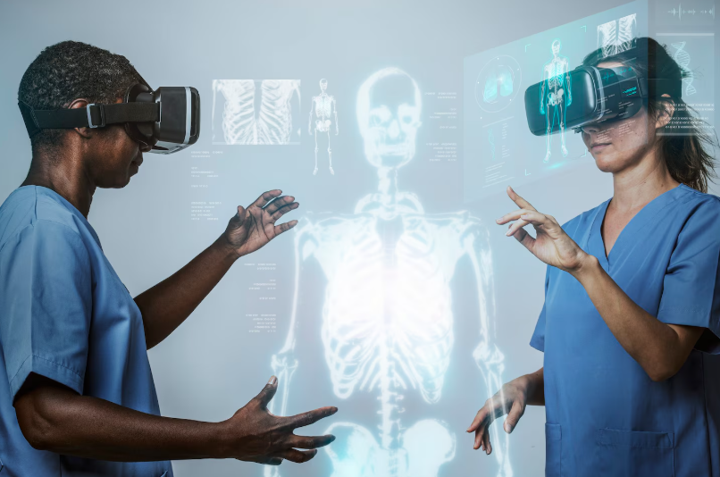
Da Vinci robotic surgery in Bangalore for cancer is performed by Prof. Dr. Sandeep Nayak at associated hospitals in Bangalore where the da Vinci system is available with his team and the consultation, treatment planning and follow-up centred at MACS Clinic in Jayanagar, and the reason that matters is that the da Vinci system on its own doesn’t produce outcomes, the surgeon operating it does and Dr. Nayak has been doing this for over 15 years at genuine volume.
According to Prof. Dr. Sandeep Nayak,Surgical Oncologist in India, “The da Vinci system is a tool and like any tool what it produces depends entirely on the experience and judgment of the person using it and a surgeon who has operated with it on your specific cancer type hundreds of times is a fundamentally different proposition from one who has done dozens.”
Where Is Da Vinci Robotic Surgery for Cancer Available in Bangalore?
These are the key things to understand about da Vinci robotic surgery availability in Bangalore:
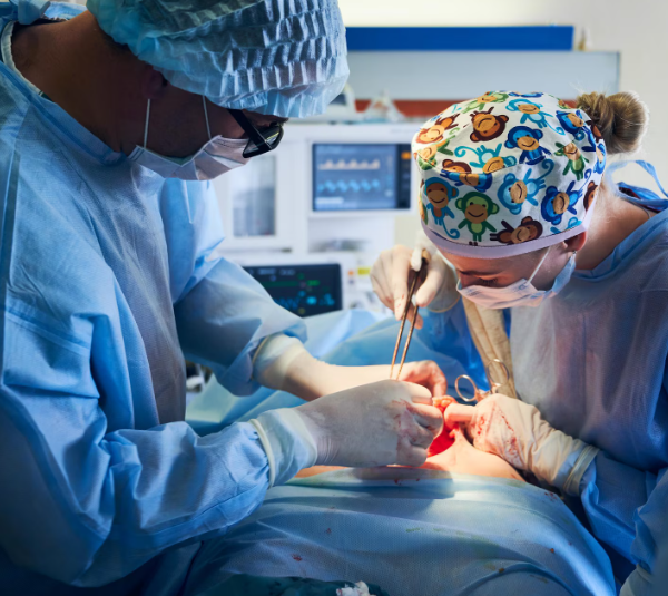
Multiple hospitals have it: Several hospitals in Bangalore now have da Vinci robotic systems but having the equipment and having a surgical oncologist who operates with it at real volume across the full cancer spectrum are two different things and the second one is what actually determines outcomes.
Dr. Nayak’s programme: Prof. Dr. Sandeep Nayak operates with the da Vinci system at associated hospitals in Bangalore with over 15 years of robotic surgical oncology experience and over a thousand robotic cancer surgeries performed making his programme the highest volume dedicated robotic cancer surgery practice in Bangalore.
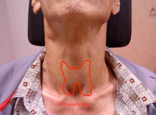
RABIT with da Vinci: RABIT scarless thyroid surgery uses the da Vinci system to navigate instruments through a tunnel from armpit and infraclavicular incisions to the thyroid in the neck and Dr. Nayak developed this technique himself specifically for the da Vinci platform making him the most credible person in India to perform it.
MIND and RIA-MIND: These robotic pelvic cancer dissection techniques developed by Dr. Nayak use the da Vinci system’s magnified 3D view and wristed instruments to operate in the narrow pelvis with precision that open surgery and standard laparoscopic surgery in that space cannot replicate and they’re available only where Dr. Nayak operates.
The hospitals in Bangalore with da Vinci robotic systems are not all equivalent for cancer surgery because the surgical programme around the system, the volume of cancer cases it’s used for and the experience of the surgeon using it vary enormously between them.Robotic cancer surgery at MACS Clinic through Dr. Nayak’s associated hospital programme is where the da Vinci system gets used by a surgeon whose robotic oncology experience in Bangalore is genuinely in a different category from most alternatives.
What Cancers Does Dr. Nayak Treat With Da Vinci Robotic Surgery in Bangalore?
These are the cancer types Dr. Nayak operates on using the da Vinci system:
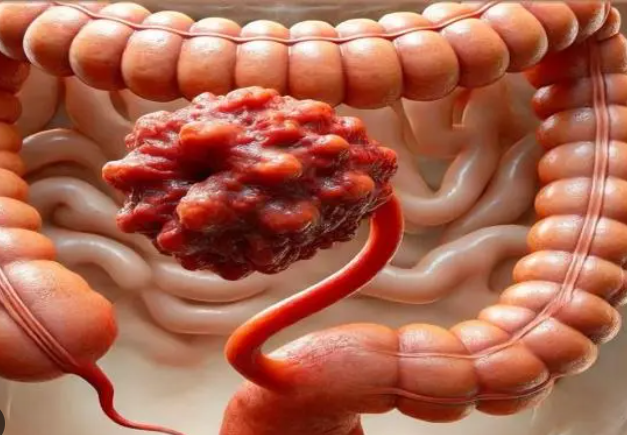
Colorectal cancer: Da Vinci robotic colon and rectal cancer surgery including D3 resection, low anterior resection and inter-sphincteric resection using MIND and RIA-MIND where the system’s capabilities in the narrow pelvic space change what sphincter preservation and nerve sparing outcomes actually look like.
Thyroid cancer: RABIT uses the da Vinci system to perform scarless thyroid surgery through armpit and infraclavicular incisions and conventional robotic thyroidectomy with neck dissection for cases where RABIT isn’t appropriate, both performed at a volume that gives Dr. Nayak specific knowledge of which fits each patient.
Urological cancers: Robotic prostatectomy and robotic kidney cancer surgery using the da Vinci system where the precision in retroperitoneal and pelvic spaces changes nerve preservation and functional outcomes compared to open or conventional laparoscopic approaches in the same anatomical territory.
Gynaecological cancers: Da Vinci robotic ovarian cancer surgery, robotic hysterectomy for uterine cancer and other gynaecological malignancies treated robotically at MACS Clinic where pelvic precision and function preservation are both part of what the surgical approach aims for.
Whether da Vinci robotic surgery is the right approach for your specific cancer, stage and anatomy is a question that needs your imaging and a surgeon whose da Vinci oncology volume gives him specific answers rather than general reassurance that the system is available.Laparoscopic cancer surgery at MACS Clinic covers the full minimally invasive spectrum alongside da Vinci robotic surgery where the approach gets matched to your case.
Why Choose Dr. Sandeep Nayak for Cancer Treatment?
Over 15 years with the da Vinci system for cancer surgery in Bangalore before most hospitals in the city had committed to the platform. Over a thousand robotic cancer surgeries. RABIT built on da Vinci. MIND built on da Vinci. RIA-MIND built on da Vinci. 24 years in surgical oncology. Chairman of Oncology Services Karnataka. Kidwai Memorial Institute of Oncology alumnus. MACS Clinic Jayanagar Bangalore for consultations, Monday to Saturday 3pm to 6:30pm, contact plus 91 9482202240. The da Vinci system is widely available in Bangalore now. A surgeon who has been using it for cancer for 15 years at this volume is not.
Frequently Asked Questions
Which hospital offers da Vinci robotic surgery in Bangalore? Prof. Dr. Sandeep Nayak performs da Vinci robotic cancer surgery at associated hospitals in Bangalore through MACS Clinic Jayanagar with over 15 years of robotic oncology experience, contact plus 91 9482202240.
What cancers does Dr. Sandeep Nayak treat with da Vinci robotic surgery? Colorectal, thyroid, prostate, kidney, ovarian and other solid tumour cancers are all treated using the da Vinci system as part of Dr. Nayak’s robotic cancer surgery programme in Bangalore.
Is da Vinci robotic cancer surgery safe in Bangalore? Yes for the right patients and procedures at a centre with genuine robotic oncology volume and da Vinci experience across multiple cancer types produces outcomes that consistently match or exceed open surgery.
How do I book a da Vinci robotic cancer surgery consultation in Bangalore? MACS Clinic Jayanagar Bangalore, Monday to Saturday 3pm to 6:30pm, contact plus 91 9482202240, bring all staging scans and biopsy reports to the first consultation.
Prof. Dr. Sandeep Nayak at MACS Clinic in Jayanagar Bangalore is where patients who’ve been told a permanent stoma is their only option for rectal cancer and haven’t accepted that answer tend to end up because his MIND and RIA-MIND techniques for robotic pelvic surgery, his volume of inter-sphincteric resection and low anterior resection and his record of sphincter preservation in cases where less experienced teams defaulted to a bag makes him the most credible answer to this specific question in Bangalore.
According to Prof. Dr. Sandeep Nayak,Surgical Oncologist in India, “A permanent stoma is not inevitable for most rectal cancer patients and the difference between who gets one and who doesn’t is often less about tumour biology and more about the surgical team’s experience with sphincter-preserving techniques in the narrow pelvis.”
What Makes Dr. Sandeep Nayak the Best Rectal Cancer Surgeon for Stoma Avoidance in Bangalore?
These are the things that put Dr. Nayak’s sphincter-preserving rectal cancer practice ahead of most alternatives in Bangalore:
MIND technique: Dr. Nayak’s own robotic pelvic dissection approach developed from operating in the narrow pelvis on low rectal cancer patients repeatedly enough to see where standard technique was leaving sphincter preservation on the table and then building a better dissection strategy rather than accepting the permanent stoma as the inevitable outcome for those cases.
ISR volume: Inter-sphincteric resection dissecting the plane between internal and external anal sphincters to remove low rectal cancer while keeping the external sphincter intact requires operating in that specific anatomy repeatedly and Dr. Nayak’s ISR volume in Bangalore is not something most rectal cancer surgeons in the city are close to matching.
Second opinion cases: Many patients who come to MACS Clinic asking about sphincter preservation were told a permanent bag was their only option somewhere else first and a significant proportion of them find after Dr. Nayak reviews their MRI that their case was actually assessable for preservation and the answer they’d received reflected the limits of the surgical team not the limits of what was possible for their tumour.
Honest patient selection: ISR and ultra-low anterior resection are not appropriate for every low rectal cancer and Dr. Nayak tells patients when preservation isn’t safely achievable rather than attempting it in cases where the outcome would compromise either the cancer clearance or the functional result in a way that doesn’t serve the patient.
Patients from across Bangalore and from other cities in South India who come to MACS Clinic specifically about stoma avoidance for rectal cancer consistently say the consultation gave them a more specific and more honest assessment of what was possible for their case than anything they’d found before.Rectal cancer treatment at MACS Clinic covers the full low rectal cancer surgical spectrum where sphincter preservation is the goal whenever the tumour location, stage and anatomy make it safely achievable.
What Should Rectal Cancer Patients Expecting Stoma Avoidance Know at MACS Clinic?
These are the things worth understanding before your consultation at MACS Clinic about rectal cancer without permanent stoma:
MRI is the starting point: Whether your tumour sits close enough to the sphincter to make preservation genuinely challenging or far enough away that it should never have been a conversation about a permanent bag in the first place gets determined from your MRI not from a general discussion about rectal cancer surgery.
Neoadjuvant therapy first: Most low rectal cancers suitable for sphincter-preserving surgery benefit from chemoradiation before the operation to shrink the tumour and create the margin that makes ISR or ultra-low anterior resection achievable and that pre-operative treatment pathway is part of the plan from the first consultation at MACS Clinic.
Temporary stoma explained: Most sphincter-preserving low rectal operations include a temporary defunctioning stoma to protect the anastomosis while it heals and that reverses in a second smaller operation a few months later so a bag immediately after surgery is not the same as having one permanently.
Function after preservation: Keeping the sphincter anatomically is not the same as keeping normal bowel function and low anterior resection syndrome with urgency, frequency and clustering is a real outcome that Dr. Nayak discusses honestly with patients before surgery rather than after they’ve already been through it.
Whether sphincter preservation is genuinely achievable for your specific rectal cancer needs your MRI, your staging and a surgeon who has operated in that pelvic space enough times to know the honest difference between possible and wishful thinking.Colon cancer treatment at MACS Clinic covers the full colorectal surgical spectrum alongside rectal cancer where surgical thoroughness and function preservation are both part of every case.
Why Choose Dr. Sandeep Nayak for Cancer Treatment?
MIND built here for exactly this situation. RIA-MIND built when MIND wasn’t enough for the hardest cases. ISR at real volume in Bangalore. Second opinions that tell patients what their MRI actually shows rather than what a previous surgical team found it convenient to offer. 24 years in surgical oncology. Over a thousand robotic cancer surgeries. Chairman of Oncology Services Karnataka. Kidwai Memorial Institute of Oncology alumnus. MACS Clinic Jayanagar Bangalore, Monday to Saturday 3pm to 6:30pm, contact plus 91 9482202240. If stoma avoidance is possible for your rectal cancer Dr. Nayak will find it. If it isn’t he’ll tell you honestly rather than attempting something that compromises your cancer outcome.
Frequently Asked Questions
Who is the best rectal cancer surgeon in Bangalore for treatment without permanent stoma? Prof. Dr. Sandeep Nayak at MACS Clinic Jayanagar Bangalore with MIND technique, ISR at real volume and a track record of sphincter preservation in cases told elsewhere a permanent bag was inevitable, contact plus 91 9482202240.
Can rectal cancer be treated without a permanent stoma in Bangalore? Yes in many cases, tumour height, response to neoadjuvant therapy and the surgeon’s ISR volume and sphincter-preserving technique are what determine whether a permanent stoma is avoidable for your specific case.
What is the difference between a temporary and permanent stoma in rectal cancer surgery? A temporary stoma protecting the bowel join while it heals gets reversed in a second smaller operation typically three to six months later so it is not the same as a permanent one.
How do I book a rectal cancer consultation at MACS Clinic Bangalore? MACS Clinic Jayanagar Bangalore, Monday to Saturday 3pm to 6:30pm, contact plus 91 9482202240, bring your MRI and all staging scans to the first consultation.
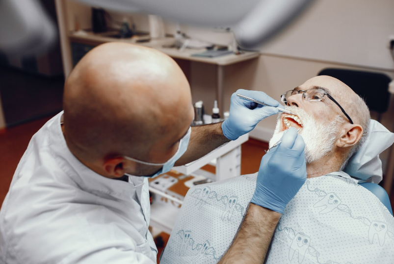
Prof. Dr. Sandeep Nayak at MACS Clinic in Jayanagar Bangalore is where patients who’ve seriously researched oral cancer surgery in Bangalore consistently end up because 24 years of surgical oncology, trans-oral robotic surgery for base of tongue and oropharyngeal cancers, neck dissection built into every appropriate operation and a practice treating the full spectrum of oral and head and neck cancers at real volume puts him in a category most oral cancer surgeons in Bangalore simply aren’t in.
According to Prof. Dr. Sandeep Nayak,Surgical Oncologist in India, “Oral cancer surgery done properly means clear margins and the right neck surgery while preserving as much speech and swallowing function as the tumour location and stage allow and getting that balance right requires operating in this anatomy repeatedly not occasionally.”
What Makes Dr. Sandeep Nayak the Best Oral Cancer Surgeon in Bangalore?
These are the things that separate Dr. Nayak’s oral cancer surgical practice from most alternatives in Bangalore:
TORS available: Trans-oral robotic surgery removes base of tongue and oropharyngeal tumours through the mouth without any external incision and the robotic camera and wristed instruments working in that confined space give access and precision that conventional open surgery through the jaw or neck genuinely cannot offer for tumours in those locations.
Full oral cancer spectrum: Tongue cancer, floor of mouth cancer, buccal mucosa cancer, lip cancer, gingival tumours, hard palate cancer, base of tongue cancer and oropharyngeal tumours all treated at MACS Clinic rather than a subset of oral cancers with the rest sent elsewhere to surgeons who may or may not have the same standard.
Neck dissection volume: Oral cancer spreads to neck lymph nodes early and reliably and Dr. Nayak’s volume of neck dissections across all oral and head and neck cancer types has built the anatomical familiarity with the neck that means clearing what needs to come out while preserving what doesn’t is something that comes from deep experience not from careful deliberation about what the anatomy textbook says.
Function preservation focus: Getting clear margins without unnecessarily sacrificing speech and swallowing function is the surgical challenge in oral cancer and it’s where the experience of a surgeon who has operated on oral cancers repeatedly at real volume shows up most directly in what patients end up with on the other side of treatment.
Patients from across Bangalore and from other cities in South India who come to MACS Clinic for oral cancer surgery consistently say the consultation gave them more clarity about their specific case, more realistic options and more honest information about what surgery would involve than they’d found anywhere else before finding Dr. Nayak.Oral cancer treatment at MACS Clinic covers the full spectrum of oral cavity and oropharyngeal cancers where surgical planning starts from your actual imaging and pathology.
What Should Oral Cancer Patients Expect at MACS Clinic Bangalore?
These are the things worth knowing before your first consultation at MACS Clinic for oral cancer:
Honest staging: Dr. Nayak reviews your imaging himself and tells you what stage your oral cancer is at, whether surgery alone covers the treatment or radiation and chemotherapy are part of the plan and what realistic outcomes look like for your specific tumour location and size.
Approach explained: Whether your oral cancer needs conventional resection, trans-oral robotic surgery or a combined approach is explained in terms of what your specific tumour location, size and pathology actually call for rather than what the centre finds most convenient or what sounds most advanced.
Reconstruction discussed: Oral cancer surgery sometimes requires reconstruction and the conversation about what that involves, what it means for speech and swallowing long term and what recovery looks like happens before surgery at MACS Clinic so patients understand the full picture of what they’re agreeing to.
Adjuvant coordination: Patients who need radiation or chemotherapy after oral cancer surgery have that transition planned before discharge from surgical care rather than being left to find and coordinate their own adjuvant treatment after Dr. Nayak has done the surgery and they’ve been sent home.
Whether MACS Clinic is the right place for your specific oral cancer depends on your staging, your tumour location and a consultation where Dr. Nayak can look at what you’ve actually got.Thyroid cancer treatment at MACS Clinic covers thyroid cancer as part of the broader head and neck cancer surgical programme where oral, oropharyngeal and thyroid cancers are all treated within the same specialist practice.
Why Choose Dr. Sandeep Nayak for Cancer Treatment?
24 years in surgical oncology. TORS for base of tongue and oropharyngeal cancers. Neck dissection at real volume across all oral and head and neck cancer types. Function preservation built into every oral cancer surgical plan. Over a thousand robotic cancer surgeries. RABIT, MIND and RIA-MIND developed here. Chairman of Oncology Services Karnataka. Kidwai Memorial Institute of Oncology alumnus. MACS Clinic Jayanagar Bangalore, Monday to Saturday 3pm to 6:30pm, contact plus 91 9482202240. Oral cancer surgery done properly gives you clear margins, the right neck clearance and the best function outcome the anatomy allows and that’s what Dr. Nayak’s practice is built around delivering.
Frequently Asked Questions
Who is the best oral cancer surgeon in Bangalore? Prof. Dr. Sandeep Nayak at MACS Clinic Jayanagar Bangalore with 24 years of surgical oncology, TORS for oral cancer and full spectrum oral cavity and oropharyngeal cancer surgery, contact plus 91 9482202240.
Does Dr. Sandeep Nayak perform trans-oral robotic surgery for oral cancer in Bangalore? Yes, TORS for base of tongue and oropharyngeal cancers is available at MACS Clinic giving patients robotic oral cancer surgery without external jaw or neck incisions.
Which oral cancers does Dr. Sandeep Nayak treat at MACS Clinic? Tongue, floor of mouth, buccal mucosa, lip, gingival, hard palate, base of tongue and oropharyngeal cancers are all treated at MACS Clinic as part of the full oral cancer surgical programme.
How do I book an oral cancer consultation at MACS Clinic Bangalore? MACS Clinic Jayanagar Bangalore, Monday to Saturday 3pm to 6:30pm, contact plus 91 9482202240, bring all imaging and biopsy reports to the first consultation.
For thyroid cancer treatment in Bangalore MACS Clinic in Jayanagar is where patients who’ve researched this properly tend to end up because Prof. Dr. Sandeep Nayak developed RABIT scarless thyroid surgery himself, has been performing robotic thyroidectomy for over 15 years in India and treats thyroid cancer at a volume and with a technique that simply isn’t available at the same level at any other centre in Bangalore.
According to Prof. Dr. Sandeep Nayak,Surgical Oncologist in India, “Thyroid cancer treatment done properly means complete surgical removal with the right neck dissection when needed and for patients who want no visible scar on their neck RABIT gives them that without compromising the oncological outcome.”
What Makes MACS Clinic the Best for Thyroid Cancer Treatment in Bangalore?
These are the things that set MACS Clinic apart for thyroid cancer treatment specifically:
RABIT developed here: Scarless thyroid surgery removing the gland through armpit and infraclavicular incisions with no cut on the neck is a technique Dr. Nayak built himself at MACS Clinic and performs at higher volume than any other centre in Bangalore or South India rather than a procedure adopted from someone else’s practice.
Volume of thyroid surgery: Total thyroidectomy, completion thyroidectomy, thyroid lobectomy and RABIT all performed at real volume means the decisions about what your specific thyroid cancer needs come from deep familiarity with the anatomy and the disease rather than from careful reference to what the guidelines say for a case the surgeon encounters occasionally.
Neck dissection integrated: Thyroid cancer spreads to neck lymph nodes and Dr. Nayak builds neck dissection into the surgical plan for every case where the staging and pathology indicate it rather than treating it as a separate procedure the patient has to go through in a second operation after the thyroid is out.
Complete pathway: Thyroid cancer treatment at MACS Clinic doesn’t end at surgery, Dr. Nayak coordinates the post-operative radioiodine planning, the TSH suppression strategy and the surveillance imaging schedule with the patient’s endocrinologist so the oncological follow-up has structure rather than being left to the patient to organise themselves after discharge.
Patients with thyroid cancer who come to MACS Clinic in Bangalore consistently say the consultation gave them a clearer picture of their specific case and more realistic options including RABIT for those who want no neck scar than they’d found anywhere else they’d been seen.Thyroid cancer treatment at MACS Clinic covers the full thyroid cancer surgical spectrum from straightforward total thyroidectomy through RABIT and complex neck dissection where the approach gets matched to what your case actually needs.
What Should Thyroid Cancer Patients Expect at MACS Clinic Bangalore?
These are the things patients coming to MACS Clinic for thyroid cancer should know before the first consultation:
Honest RABIT assessment: Not every thyroid cancer patient qualifies for RABIT and Dr. Nayak will tell you honestly whether your tumour extent, your anatomy and your fitness make it the right approach rather than offering it to every patient who asks for it because it sounds better than conventional surgery.
Staging before planning: Dr. Nayak reviews your ultrasound, your cytology and your staging scans himself before the consultation and the treatment plan that comes out of that appointment is built from what’s actually on your imaging not from a protocol applied before anyone looked at it.
Conventional when it fits better: Some thyroid cancers are better served by conventional thyroidectomy with a neck incision than by RABIT and at MACS Clinic that honest recommendation gets made rather than RABIT being pushed because it’s the more impressive sounding option or the more profitable one.
Post-operative plan included: Radioiodine ablation timing, thyroid hormone replacement dose, TSH suppression targets and surveillance intervals are discussed before surgery at MACS Clinic so the patient understands the full treatment pathway rather than finding out what comes next only after the operation is already done.
Whether MACS Clinic is the right place for your specific thyroid cancer depends on your histology, your staging and a consultation where Dr. Nayak can look at what you’ve actually got and tell you what the realistic options are.Robotic cancer surgery at MACS Clinic covers the full robotic oncology spectrum where RABIT sits alongside other robotic cancer surgery approaches as part of a comprehensive practice.
Why Choose Dr. Sandeep Nayak for Cancer Treatment?
RABIT built here not borrowed. Over 15 years of robotic thyroid surgery in India. Total thyroidectomy, neck dissection, RABIT all at real volume. Radioiodine coordination included. 24 years in surgical oncology. Over a thousand robotic cancer surgeries across cancer types. Chairman of Oncology Services Karnataka. Kidwai Memorial Institute of Oncology alumnus. MACS Clinic Jayanagar Bangalore, Monday to Saturday 3pm to 6:30pm, contact plus 91 9482202240. Thyroid cancer treated properly means complete removal, the right neck surgery and a post-operative plan that doesn’t leave the patient working it out themselves and that’s what MACS Clinic is built to deliver.
Frequently Asked Questions
What is the best hospital for thyroid cancer treatment in Bangalore? MACS Clinic Jayanagar Bangalore under Prof. Dr. Sandeep Nayak offers thyroid cancer surgery including RABIT scarless thyroidectomy and neck dissection with over 15 years of robotic thyroid surgery, contact plus 91 9482202240.
Does Dr. Sandeep Nayak perform RABIT scarless thyroid surgery in Bangalore? Yes, RABIT removes the thyroid through armpit and infraclavicular incisions with no neck scar and is available at MACS Clinic Bangalore where Dr. Nayak developed the technique himself.
How is thyroid cancer treated at MACS Clinic Bangalore? Total thyroidectomy with or without neck dissection using conventional or RABIT approach followed by radioiodine ablation and TSH suppression coordinated with the patient’s endocrinologist.
How do I book a thyroid cancer consultation at MACS Clinic Bangalore? MACS Clinic Jayanagar Bangalore, Monday to Saturday 3pm to 6:30pm, contact plus 91 9482202240, bring ultrasound, cytology and any prior treatment records to the first consultation.
Prof. Dr. Sandeep Nayak at MACS Clinic in Jayanagar Bangalore is where patients from across South India end up when they’ve asked seriously about laparoscopic cancer surgery because 15 years of robotic and laparoscopic cancer surgery before most South Indian centres committed to it, over a thousand minimally invasive cancer operations and original techniques built from operating at that volume rather than adopted from someone else’s practice is a combination that doesn’t exist at many places in the region.
According to Prof. Dr. Sandeep Nayak,Surgical Oncologist in India, “The best laparoscopic cancer surgeon is not the one with the most impressive equipment but the one who has operated laparoscopically on your specific cancer type often enough that the decisions in theatre come from deep familiarity rather than careful deliberation.”
What Makes Dr. Sandeep Nayak the Best Laparoscopic Cancer Surgeon in South India?
These are the things that put Dr. Nayak’s laparoscopic cancer surgery practice ahead of most alternatives in the region:
15 years of volume: Getting into laparoscopic cancer surgery over 15 years ago in South India before most centres had committed to it and building a practice at real volume from that early start means Dr. Nayak has operated laparoscopically on cancer cases that surgeons who adopted minimally invasive surgery more recently are still encountering for the first time.
Full cancer spectrum: Colorectal, gastric, liver, kidney, adrenal, ovarian, uterine and other cancers all done laparoscopically at MACS Clinic rather than laparoscopic surgery available for two or three cancer types with the rest done open because the surgeon’s minimally invasive experience doesn’t extend beyond a narrow range.
Original techniques: RABIT, MIND and RIA-MIND came from operating laparoscopically and robotically at real volume and paying close enough attention to outcomes to see where standard technique was leaving something on the table that a better approach could recover and then building that approach rather than accepting the limit.
D3 resection laparoscopically: Complete mesocolic excision with central vascular ligation for colon cancer done laparoscopically requires specific technical depth beyond standard laparoscopic colorectal surgery and Dr. Nayak does this as standard practice rather than as a technically demanding exception to how most of his colon cancer cases get done.
Patients from Chennai, Hyderabad, Kochi, Coimbatore, Mysore and across South India travel to MACS Clinic for laparoscopic cancer surgery specifically because the combination of volume, cancer spectrum coverage and techniques like MIND and RABIT isn’t available at the same level at any single centre closer to where they live.Laparoscopic cancer surgery at MACS Clinic covers the full minimally invasive spectrum across all these cancer types where every surgical decision starts from what your specific case needs.
Which Cancers Does Dr. Nayak Treat Laparoscopically in South India?
These are the cancer types Dr. Nayak operates on laparoscopically at MACS Clinic:
Colorectal: Colon and rectal cancer including D3 resection, complete mesocolic excision, low anterior resection and inter-sphincteric resection done laparoscopically and robotically as standard at MACS Clinic rather than the open approach reserved for cases where minimally invasive isn’t being attempted.
Gastric and upper GI: Gastric cancer, oesophageal junction tumours and upper gastrointestinal cancers done laparoscopically where the staging, location and patient fitness allow it giving patients the recovery advantage that open gastrectomy patients in South India are still waiting weeks longer to reach.
Urological and gynaecological: Kidney cancer, adrenal tumours, prostate cancer, ovarian cancer and uterine cancer all treated laparoscopically at MACS Clinic where the robotic system’s precision in the retroperitoneal and pelvic spaces changes what nerve preservation and function outcomes actually look like for patients.
Liver and hepatobiliary: Liver tumours, gallbladder cancer and bile duct cancers approached laparoscopically where the anatomy and disease extent allow it by a surgeon whose hepatic laparoscopic experience runs deep enough that the access and bleeding control in that territory is something built from volume rather than approached cautiously case by case.
Whether laparoscopic surgery fits your specific cancer type, stage and anatomy is a question a consultation at MACS Clinic answers specifically rather than generally because the honest answer varies with what’s actually on your scans.Robotic cancer surgery at MACS Clinic sits alongside laparoscopic surgery as part of the full minimally invasive spectrum where the right approach gets matched to the case.
Why Choose Dr. Sandeep Nayak for Cancer Treatment?
Colorectal. Gastric. Kidney. Liver. Prostate. Ovarian. All of them laparoscopically for 15 years before most South Indian centres got there. RABIT built here. MIND built here. RIA-MIND built here. D3 resection laparoscopically as standard not as an exception. Over a thousand minimally invasive cancer surgeries. Chairman of Oncology Services Karnataka. Kidwai Memorial Institute of Oncology alumnus. MACS Clinic Jayanagar Bangalore, Monday to Saturday 3pm to 6:30pm, contact plus 91 9482202240. Patients across South India who ask the right questions about laparoscopic cancer surgery end up at the same place.
Frequently Asked Questions
Who is the best laparoscopic cancer surgeon in South India? Prof. Dr. Sandeep Nayak at MACS Clinic Jayanagar Bangalore with 15 years of laparoscopic cancer surgery, full cancer spectrum coverage and original techniques RABIT, MIND and RIA-MIND, contact plus 91 9482202240.
Which cancers does Dr. Sandeep Nayak treat laparoscopically in South India? Colorectal, gastric, kidney, liver, adrenal, prostate, ovarian and uterine cancers among others are all treated laparoscopically at MACS Clinic Bangalore.
Does Dr. Sandeep Nayak perform D3 laparoscopic resection for colon cancer? Yes, D3 resection with complete mesocolic excision done laparoscopically is standard practice at MACS Clinic rather than an exception reserved for selected cases.
How do I book a laparoscopic cancer surgery consultation at MACS Clinic? MACS Clinic Jayanagar Bangalore, Monday to Saturday 3pm to 6:30pm, contact plus 91 9482202240, bring all staging scans and biopsy reports to the first consultation.
Dr. Sandeep Nayak, a board-certified surgical oncologist (M.ch ., D.N.B.), specializes in minimally invasive cancer surgery, including robotic and laparoscopic techniques.