
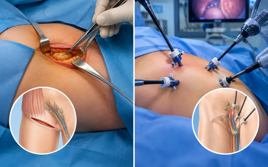
When Is a Second Cancer Surgery Needed?

When Is a Second Cancer Surgery Needed?

A second cancer surgery is needed when the first operation didn’t fully finish the job, or when the cancer comes back. The most common reason is a positive margin, where pathology shows cancer at the edge of what was removed. Other triggers are recurrence, an incidental cancer found in the specimen, or upstaging after pathology. The final report usually decides it.

According to Dr. Sandeep Nayak, Surgical Oncologist in India, “Nobody wants to hear they need a second operation, but sometimes the pathology leaves no choice. The commonest reason is a positive margin, cancer right at the cut edge, which means some may have been left behind. We go back to clear it. Other times the cancer returns, or the final report shows the disease was more than we expected. The report guides the decision, not guesswork.”

Waiting on a pathology result and worried about more surgery?

What Are the Main Reasons?

A handful of clear situations call for a return to theatre.

  • Positive margins : The biggest reason. If cancer reaches the edge of the removed tissue, a re-excision takes more to be sure it’s all gone.
  • Recurrence : Cancer that comes back in the same area, after the first surgery and any treatment, often needs a second operation to remove it.
  • Incidental cancer : Sometimes cancer is found by surprise in a removed organ, like a gallbladder taken for stones. That can need a wider second surgery.
  • Upstaging : When the final pathology shows the disease was more advanced than thought, a more extensive operation may be needed to match it.

The single biggest trigger ties directly to the robotic cancer surgery precision of the first operation, since a clean first removal is what avoids a second.

How Is a Second Surgery Avoided?

The best way to avoid a repeat operation is to get the first one right.

  • Clear margins first time : A complete removal with a healthy rim of tissue is the goal. Achieve that, and a second surgery usually isn’t needed.
  • Good imaging : Accurate scans before surgery map the tumour properly, so the surgeon knows exactly how much to take. Less guesswork, fewer surprises.
  • Intraoperative checks : Tools like frozen section and intraoperative ultrasound let the surgeon confirm clearance during the operation itself.
  • Experience : A high volume surgeon judges the right amount to remove first time. That skill is what keeps the re-operation rate low.

When a second surgery is for spread rather than margins, understanding metastatic cancer explains why removing returned or isolated disease can still offer a real benefit.

Why Choose Dr. Sandeep Nayak for Cancer Surgery?

Dr. Sandeep Nayak is a surgical oncologist with 24 years behind him and a fellowship in laparoscopic and robotic onco-surgery. His focus on precise first time surgery, clear margins, accurate staging, careful imaging, is what keeps second operations to a minimum. When a second surgery genuinely is needed, that same experience guides it, whether it’s a re-excision, removing a recurrence, or handling an incidental finding. Getting it right matters more the second time, not less.

A second operation is harder than the first. Scar tissue, altered anatomy and a patient who’s already been through one surgery all raise the stakes. This is exactly where a high volume surgeon earns their place, judging when a second surgery will genuinely help and executing it cleanly when it will. The goal is always to make the first operation complete, and to handle the second with the skill it demands when it can’t be avoided.

Frequently Asked Questions

When is a second cancer surgery needed?

For positive margins, recurrence, an incidental cancer, or upstaging found after pathology.

What is re-excision surgery?

A second operation to remove more tissue when cancer reaches the specimen edge.

Does positive margin always mean more surgery?

Usually, unless the repeat surgery’s risks outweigh its benefit for that patient.

Can a second surgery be avoided?

Often, when the first surgery achieves clear margins and removes the cancer completely.

References

  1. Re-excision after positive margins in breast surgery — National Library of Medicine
  2. Predictors of re-excision following breast-conserving surgery — National Library of Medicine

Disclaimer: This blog is for informational and educational purposes only and is not a substitute for professional medical advice or diagnosis.

Can Laparoscopy Be Used for Liver Cancer?

Can Laparoscopy Be Used for Liver Cancer?

Laparoscopy can be used for liver cancer, in the right patient. For small tumours sitting in accessible parts of the liver, keyhole surgery removes them with the same cancer outcomes as open surgery, plus a faster, gentler recovery. It isn’t suited to every case. Large tumours, or ones wrapped around major blood vessels, still call for open surgery. Tumour size and position decide it.

According to Dr. Sandeep Nayak, Surgical Oncologist in India, “Laparoscopic liver surgery has come a long way, and for the right tumour it’s an excellent option. A small cancer in an accessible segment comes out cleanly through keyhole incisions, and the patient recovers far quicker. The survival and margins match open surgery. But I won’t force it. A big central tumour near the main vessels is safer done open. The case decides the approach.”

Wondering if your liver tumour can be removed by keyhole surgery?

When Does Laparoscopy Work for Liver Cancer?

Keyhole liver surgery suits specific tumours, and selecting them well is the key.

  • Small tumours : A small, contained cancer is the ideal candidate. The smaller and more defined it is, the better suited to a keyhole approach.
  • Peripheral location : Tumours in the outer, more accessible parts of the liver are far easier to reach laparoscopically than deep central ones.
  • Away from vessels : A tumour clear of the major blood vessels can be removed safely. Proximity to those vessels is what tips toward open surgery.
  • Good liver function : The patient’s liver needs enough healthy reserve, especially where cirrhosis is in the picture, to handle the resection.

This precision is the foundation of modern robotic cancer surgery, where the same minimally invasive principles apply to complex liver work.

Why Choose It Over Open Surgery?

When a tumour suits the keyhole route, the advantages for the patient are real.

  • Less blood loss : Laparoscopic liver surgery typically means less bleeding during the operation. That matters a great deal in liver work.
  • Faster recovery : Smaller incisions mean less pain and a quicker return home. Patients are often up and about much sooner.
  • Same cancer control : This is the crucial part. Survival, clear margins and recurrence rates match open surgery in suitable cases.
  • Earlier next steps : A faster recovery means any chemotherapy needed afterward can start sooner, which can matter for the overall outcome.

Whether surgery offers a cure at all depends on the stage, which is covered in our guide on liver cancer and when it can be treated successfully.

Why Choose Dr. Sandeep Nayak for Liver Cancer Surgery?

Dr. Sandeep Nayak is a surgical oncologist with 24 years behind him and a fellowship in laparoscopic and robotic onco-surgery. He performs minimally invasive liver resections for suitable patients, choosing the keyhole route where it genuinely helps and open surgery where safety demands it. The approach starts with honest case selection, since liver surgery punishes overreach, and the right tumour for laparoscopy is a specific thing. That judgement is what makes the technique safe.

Liver surgery is among the most demanding work in oncology, and the minimally invasive version more so. Reading the imaging, judging the tumour’s relationship to the vessels, and knowing when to switch to open is what separates a good liver surgeon from a risky one. For the right patient, laparoscopic resection offers a cure with a recovery that open surgery simply can’t match. Matching the method to the tumour is the whole craft.

Frequently Asked Questions

Can laparoscopy be used for liver cancer?

Yes. Selected liver cancers can be removed laparoscopically with outcomes equal to open surgery.

Which liver tumours suit laparoscopic surgery?

Small, peripheral, unilobar tumours away from major blood vessels suit it best.

Is laparoscopic liver surgery as effective as open?

Yes. Survival, margins and recurrence match open surgery in suitable patients.

When is open liver surgery still needed?

For large, central tumours or those involving major blood vessels, open surgery is safer.

References

  1. Minimally invasive liver surgery for hepatocellular carcinoma — National Library of Medicine
  2. Minimally invasive liver resection for colorectal metastases — National Library of Medicine

Disclaimer: This blog is for informational and educational purposes only and is not a substitute for professional medical advice or diagnosis.

Bile Duct Surgery vs Stenting: Which Is Better?

Bile Duct Surgery vs Stenting: Which Is Better?

It depends on whether cure is the goal. Surgery is the only option that can remove the cancer and offer a cure, used when the tumour is resectable and the patient is fit. Stenting doesn’t remove anything. It reopens the blocked duct to relieve jaundice, used when surgery isn’t possible or as a bridge before it. They serve different purposes, not the same one.

According to Dr. Sandeep Nayak, Surgical Oncologist in India, “These two aren’t really rivals, they answer different questions. If the tumour can be removed and the patient can withstand the operation, surgery is the path to a cure. A stent never cures anything. It clears the jaundice and makes a patient comfortable, which matters enormously when surgery isn’t on the table. Sometimes we even stent first, then operate. The decision rests on whether cure is achievable.”

Facing a blocked bile duct and weighing the options?

When Is Surgery the Right Choice?

Surgery is the answer when the goal is to remove the cancer entirely.

  • Curative intent : Only surgery can remove the tumour and offer a real chance at cure. For a resectable cancer, that makes it the first choice.
  • Resectable tumour : The cancer has to be removable, confined enough that a surgeon can take it out with clear margins. Imaging decides this.
  • Fit patient : Bile duct surgery is major, often a Whipple operation. The patient needs to be well enough to come through it.
  • Long term gain : When it works, surgery changes the whole trajectory. Stenting alone never offers that kind of outcome.

For resectable disease in a fit patient, the right bile duct cancer plan centres on surgery, with stenting playing only a supporting role.

Surgery or Stenting: How Do They Compare?

Here’s how the two line up side by side.

Feature

Surgery

Stenting

Goal

Cure

Relieve jaundice

Removes cancer

Yes

No

Best for

Resectable, fit

Unresectable, unfit

Invasiveness

Major operation

Minimal

Recovery

Weeks

Quick

As a bridge

The destination

Before surgery

  • Different goals : Surgery aims to cure. Stenting aims to comfort and decompress. Judging which one a patient needs starts with that distinction.
  • Stenting’s role : When a tumour can’t be removed, a stent restores bile flow, lifts the jaundice and lets a patient feel human again.
  • The bridge use : A stent can relieve severe jaundice first, stabilising a patient before the bigger curative surgery is done.
  • Allowing chemo : Clearing the jaundice with a stent also lets a patient start systemic chemotherapy that high bilirubin would otherwise block.

This is part of the wider scope of bile duct surgery within hepatobiliary cancer care, where surgical and palliative tools each have their place.

Why Choose Dr. Sandeep Nayak for Bile Duct Cancer Care?

Dr. Sandeep Nayak is a surgical oncologist with 24 years behind him and a fellowship in laparoscopic and robotic onco-surgery. He treats hepatobiliary cancers, including complex bile duct resections, where judging resectability correctly decides whether a patient gets a shot at cure or the right palliative path. The work starts with honest staging, since offering surgery where it can’t help, or stenting where surgery could cure, both fail the patient. That judgement is the core of it.

Bile duct cancer is unforgiving of the wrong call. A resectable tumour managed with a stent alone loses a curative chance that won’t come back. An unresectable one pushed into surgery puts a patient through a major operation for nothing. Reading the imaging accurately, staging honestly, and matching the tool to the situation is what separates good hepatobiliary care from guesswork.

Frequently Asked Questions

Is bile duct surgery better than stenting?

Surgery offers a cure when the tumour is resectable. Stenting only relieves the blockage.

What does a bile duct stent do?

It reopens a blocked duct to relieve jaundice and itching, but doesn’t remove cancer.

When is stenting chosen over surgery?

When the tumour is unresectable, the patient is unfit, or before planned surgery.

Can a stent be used before surgery?

Yes. A stent can relieve jaundice first, before definitive surgery is performed later.

References

  1. Percutaneous biliary stenting in malignant obstruction — National Library of Medicine
  2. Obstructive jaundice diagnosis and management — National Library of Medicine

Disclaimer: This blog is for informational and educational purposes only and is not a substitute for professional medical advice or diagnosis.
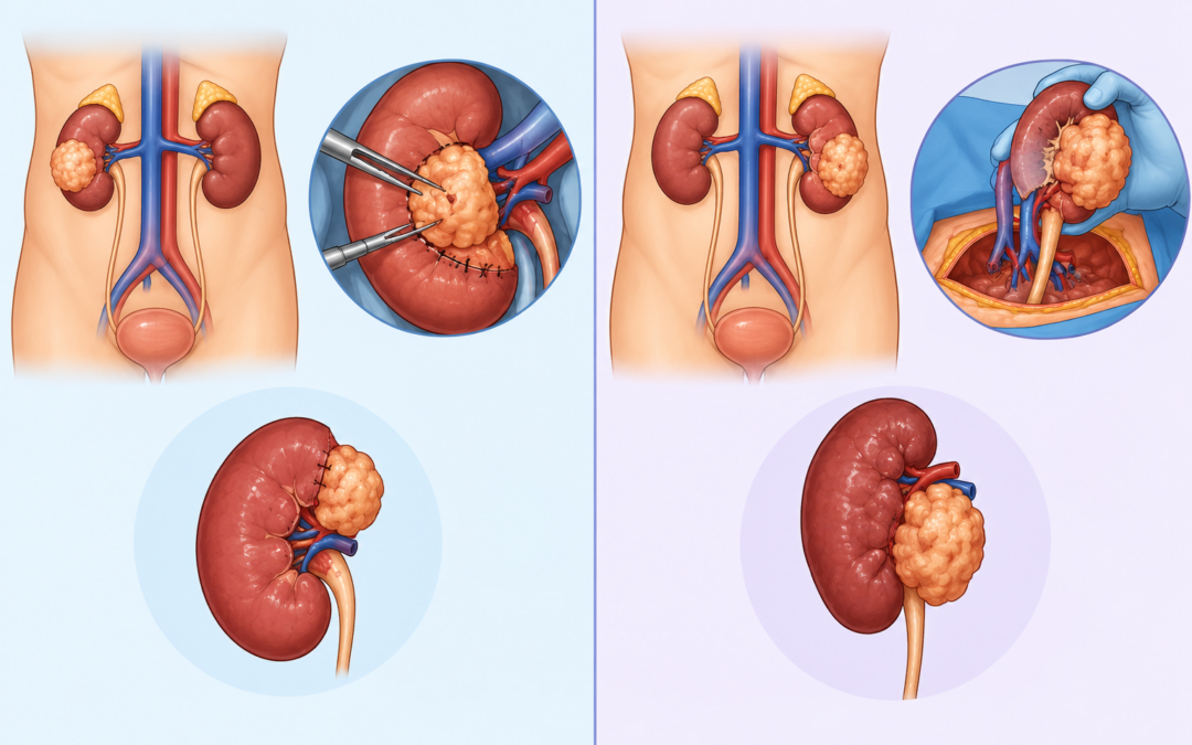
Is Appendix Cancer Treated Like Colon Cancer?

Is Appendix Cancer Treated Like Colon Cancer?

The treatment depends entirely on the tumour type. Higher grade adenocarcinomas of the appendix are treated much like colon cancer, with a right hemicolectomy and lymph node clearance. The common mucinous types are different. They spread across the abdomen as pseudomyxoma peritonei and need cytoreductive surgery with HIPEC, not standard colon cancer treatment. The histology sets the path. 

According to Dr. Sandeep Nayak, Surgical Oncologist in India, “People assume appendix cancer is just a type of colon cancer, but that’s only half right. The aggressive adenocarcinomas, yes, we often treat those like colon cancer. But the mucinous tumours are a different beast. They produce jelly like material and spread across the peritoneum. Those need HIPEC, not colon chemo. Getting the histology right is what decides the whole plan.”

Diagnosed with an appendix tumour and unsure of the path?

When Is It Treated Like Colon Cancer?

For certain appendix tumours, the colon cancer playbook genuinely applies.

  • Adenocarcinoma : Higher grade appendiceal adenocarcinomas behave like colon cancer. A right hemicolectomy to remove the appendix, nearby colon and lymph nodes is standard.
  • Shared biology : These tumours arise from similar cells to colon cancer, so the surgical logic and lymph node clearance carry across.
  • Chemo overlap : When chemotherapy is needed, the regimens often mirror those used for colon cancer. The drugs are familiar territory.
  • Staging similar : Staging follows comparable principles, looking at how deep the tumour goes and whether it’s reached the nodes.

For these cases the surgical approach overlaps heavily, though specialised HIPEC treatment enters the picture the moment the tumour spreads across the peritoneum.

When Is It Treated Completely Differently?

The mucinous appendix tumours follow a path colon cancer treatment simply doesn’t cover.

  • Mucinous neoplasms : Low grade mucinous tumours produce jelly like mucin. They rarely spread through blood or nodes the way colon cancer does.
  • Pseudomyxoma peritonei : When these rupture, mucin spreads across the abdomen. This condition needs a very different, specialised approach.
  • CRS and HIPEC : The treatment is cytoreductive surgery to remove all visible disease, then heated chemo washed through the abdomen. Not colon chemo.
  • Strong outcomes : For these tumours, CRS with HIPEC has pushed five year survival far higher than systemic chemotherapy ever achieved. A genuine shift.

This is exactly the territory covered by cytoreductive surgery and HIPEC, where appendiceal tumours are among the cancers it treats most successfully.

Why Choose Dr. Sandeep Nayak for Appendix Cancer Care?

Dr. Sandeep Nayak is a surgical oncologist with 24 years behind him and a fellowship in laparoscopic and robotic onco-surgery. He’s among India’s most experienced HIPEC surgeons, treating appendiceal tumours and pseudomyxoma peritonei alongside colorectal and ovarian peritoneal disease. The work begins with reading the histology correctly, since an appendix cancer treated as plain colon cancer, when it’s actually mucinous, misses the right treatment entirely. That distinction is where expertise shows.

Appendix cancer is where the wrong assumption costs the most. A mucinous tumour handled with standard colon cancer chemo will progress, because those tumours barely respond to it. The right answer is aggressive surgery and HIPEC, in experienced hands. Matching the treatment to the actual tumour type, rather than the organ it came from, is what gives these patients their real chance.

Frequently Asked Questions

Is appendix cancer treated like colon cancer?

Sometimes. It depends on the tumour type, since many appendix cancers need different treatment.

When is appendix cancer treated like colon cancer?

Higher grade adenocarcinomas often need a right hemicolectomy, much like colon cancer.

How are mucinous appendix tumours treated?

They often spread as pseudomyxoma peritonei, treated with cytoreductive surgery and HIPEC.

Why does the tumour type matter so much?

Because grade and spread decide whether standard colon surgery or HIPEC is needed.

References

  1. CRS and HIPEC for appendiceal pseudomyxoma peritonei survival — National Library of Medicine
  2. Appendiceal mucinous neoplasm and pseudomyxoma peritonei — National Library of Medicine

Disclaimer: This blog is for informational and educational purposes only and is not a substitute for professional medical advice or diagnosis.
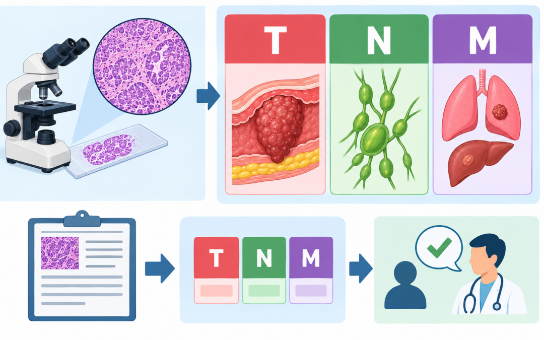
Colonoscopy vs CT Colonography: Which Is Better?

Colonoscopy vs CT Colonography: Which Is Better?

Colonoscopy is the better test. It does two jobs in one sitting, it finds polyps and removes them on the spot. CT colonography only detects. It’s a scan that locates polyps but can’t remove them, so anything it finds still needs a colonoscopy afterward. Colonoscopy screens and treats together. CT colonography screens only.

According to Dr. Sandeep Nayak, Surgical Oncologist in India, “Colonoscopy is the gold standard for a simple reason, it finds and fixes in one go. We see a polyp, we remove it, and that polyp never becomes cancer. CT colonography is useful when a colonoscopy can’t be completed or isn’t safe for someone. But a positive scan still ends in a colonoscopy. So for most people, going straight to the real thing makes sense.”

Due for a colon screening and unsure which test?

What Sets the Two Apart?

They examine the same colon but work in fundamentally different ways.

  • Colonoscopy : A flexible camera goes in and the doctor sees the colon directly. Spot a polyp, remove it then and there. One and done.
  • CT colonography : A CT scanner builds detailed images of the colon from outside. It’s often called virtual colonoscopy. It detects, but it can’t remove.
  • The big gap : Colonoscopy treats as it screens. CT colonography only reports. Anything it flags still sends you for a colonoscopy anyway.
  • Comfort tradeoff : CT colonography is less invasive and needs no sedation. But it uses radiation, and the bowel prep is much the same either way.

Catching disease early is the goal of both, and the right colon cancer treatment starts with whichever test fits the individual best.

Colonoscopy or CT Colonography: How Do They Compare?

Here’s how the two line up side by side.

Feature

Colonoscopy

CT Colonography

Removes polyps

Yes

No

Sedation

Usually

None

Radiation

None

Yes

Detects small polyps

Excellent

Can miss some

Follow up needed

Rarely

If positive

Best as

Primary test

Alternative

  • Sensitivity : Colonoscopy catches small and flat polyps that a scan can miss. For thorough detection, the camera still wins.
  • The one stop benefit : Only colonoscopy turns screening into treatment. Removing a polyp during the test prevents the cancer outright.
  • When scans help : CT colonography suits patients who can’t have a full colonoscopy, or where one couldn’t reach the whole colon.
  • The catch with CT : A clear scan is reassuring, but a positive one means a second procedure. Two preps, two appointments, more time.

Understanding early detection of colorectal cancer shows why catching polyps early, and removing them, changes the whole picture.

Why Choose Dr. Sandeep Nayak for Colon Cancer Care?

Dr. Sandeep Nayak is a surgical oncologist with 24 years behind him and a fellowship in laparoscopic and robotic onco-surgery. His colon cancer work is known internationally, particularly D3 resection, and he sees screening as the front line that prevents surgery in the first place. The approach matches the test to the patient, recommending colonoscopy where it fits and CT colonography where a full scope isn’t possible. That judgement is what makes screening genuinely useful.

The screening choice sets up everything that follows. A colonoscopy that finds and removes a polyp can stop a cancer before it ever forms, which is the cheapest, simplest win in all of oncology. When that isn’t possible, knowing the alternatives and their limits matters. Picking the right test for the right person is the quiet step that prevents far bigger problems down the line.

Frequently Asked Questions

Is colonoscopy better than CT colonography?

Colonoscopy is the gold standard since it both detects and removes polyps together.

What is CT colonography?

A CT scan that creates detailed images of the colon to look for polyps.

Does CT colonography need a follow up colonoscopy?

Yes. If it finds a polyp, a colonoscopy is still needed to remove it.

When is CT colonography preferred?

When colonoscopy is unsafe, incomplete or a patient can’t tolerate the procedure.

References

  1. Colonoscopy versus CT colonography screening trial — National Library of Medicine
  2. CT colonography as a triage technique in screening — National Library of Medicine

Disclaimer: This blog is for informational and educational purposes only and is not a substitute for professional medical advice or diagnosis.

Can HIPEC Treat Stage 4 Ovarian Cancer?

Can HIPEC Treat Stage 4 Ovarian Cancer?

HIPEC can help some Stage 4 ovarian cancer patients, but not all. It works when the cancer has spread within the abdomen and a surgeon can remove all visible disease. It isn’t a blanket Stage 4 treatment. The deciding factors are where exactly the cancer has spread and whether complete removal is achievable. Patient selection is everything here.

According to Dr. Sandeep Nayak, Surgical Oncologist in India, “HIPEC isn’t a magic answer for every Stage 4 ovarian cancer, and anyone who says so is overselling it. Where it genuinely helps is when the disease sits inside the abdomen and I can remove all of it surgically. Then the heated chemo handles what’s left behind. But if cancer has spread to the lungs or deep into the liver, HIPEC isn’t the right tool. Selecting the right patient is the whole skill.”

Want to know if HIPEC is an option in your case?

When Can HIPEC Help in Stage 4?

The treatment works in a specific situation, and getting that situation right is the key.

  • Abdominal spread : HIPEC suits cancer that’s spread across the peritoneum but stayed inside the abdomen. That’s the disease pattern it targets.
  • Complete removal : It only works if surgery can clear all visible tumour first. Leftover disease means the heated chemo has too much to handle.
  • The PCI score : Surgeons score the spread from 1 to 39. A reasonable score means HIPEC is worth offering. Too high, and it isn’t.
  • Good enough fitness : This is major surgery, often 5 to 12 hours. The patient has to be fit enough to come through it well.

This careful selection is what makes HIPEC treatment effective rather than just aggressive, and it’s why not every patient is a candidate.

What Does It Actually Involve?

HIPEC is really two procedures working together, done in a single operation.

  • Cytoreduction first : The surgeon removes every visible tumour from the abdomen. This part is the heavy lifting, and its completeness drives the outcome.
  • Heated chemo : Warmed chemotherapy is then washed through the abdominal cavity. The heat helps it penetrate and kill microscopic disease surgery can’t see.
  • The survival data : In selected patients, CRS with HIPEC has shown real survival gains. It’s not a cure, but it can buy meaningful time.
  • The recovery : It’s a big operation. A hospital stay of 10 to 14 days and a couple of months of recovery is the realistic picture.

The role of CRS and HIPEC in ovarian cancer surgery is set out in detail, including where it fits among the other treatment options.

Why Choose Dr. Sandeep Nayak for HIPEC?

Dr. Sandeep Nayak is a surgical oncologist with 24 years behind him and a fellowship in laparoscopic and robotic onco-surgery. He’s among India’s most experienced HIPEC surgeons, treating peritoneal spread from ovarian, colorectal, appendix and other cancers. The work begins with honest selection, scoring the spread and judging whether complete cytoreduction is realistic, because HIPEC only helps when the disease and the patient genuinely fit it. That judgement is where the real expertise lies.

Ovarian cancer is where HIPEC has some of its strongest support, but only in the right hands and the right patient. The completeness of the surgery is the single biggest factor in how well someone does, and that comes down to surgical skill in the abdomen. For a carefully chosen Stage 4 patient, this combination offers something that systemic chemotherapy alone often can’t.

Frequently Asked Questions

Can HIPEC treat Stage 4 ovarian cancer?

It can help selected Stage 4 patients when disease stays inside the abdomen.

Who qualifies for HIPEC in ovarian cancer?

Patients whose tumour can be fully removed and whose PCI score is within limits.

Is HIPEC a cure for ovarian cancer?

Not a cure, but it can improve survival when combined with complete surgery.

When is HIPEC not suitable?

When cancer has spread outside the abdomen or complete tumour removal isn’t possible.

References

  1. CRS plus HIPEC in advanced ovarian cancer meta-analysis — National Library of Medicine
  2. HIPEC in primary and recurrent ovarian cancer review — National Library of Medicine

Disclaimer: This blog is for informational and educational purposes only and is not a substitute for professional medical advice or diagnosis.

Call Now Button