What Is Pheochromocytoma and How Is It Diagnosed?

 What Is Pheochromocytoma and How Is It Diagnosed?

A pheochromocytoma is a rare tumour of the adrenal gland that pumps out adrenaline and related hormones. That flood of hormones drives spells of high blood pressure, headache, sweating and a pounding heart. It’s usually benign. Diagnosis starts with a blood or urine test measuring metanephrines, the breakdown products of those hormones, then a scan to locate the tumour. Getting it right before any surgery is critical.

According to Dr. Sandeep Nayak, Surgical Oncologist in India, “A pheochromocytoma is uncommon, but it’s one tumour you never want to miss. It floods the body with adrenaline, causing those dramatic spells of high blood pressure and palpitations. We diagnose it by measuring metanephrines first, then imaging to find it. The reason this matters so much is safety. Operating on an unprepared pheochromocytoma can trigger a dangerous crisis, so the diagnosis has to come first, every time.”

Having unexplained blood pressure spells and palpitations?

What Is It and What Does It Do?

Understanding this tumour means understanding the hormones it releases and the havoc they cause.

  • Where it sits : It arises in the medulla, the inner part of the adrenal gland, which normally produces adrenaline in controlled amounts. This tumour makes far too much.
  • The hormone surge : It pours out catecholamines, adrenaline and noradrenaline, unchecked. That surge is behind every symptom the tumour causes.
  • The classic triad : Headache, sweating and palpitations occurring together, often in episodes, is the hallmark. Anxiety, tremor and pallor often join them.
  • Blood pressure spells : The signature feature is high blood pressure, sometimes constant, often in sudden dramatic spikes that come and go without obvious cause.

This is one of the more demanding tumours handled within adrenal tumor treatment, precisely because of the hormones it releases.

How Is It Diagnosed?

The diagnosis follows a careful order, hormones first, then imaging, for good reason.

  • Metanephrine testing : The key test measures metanephrines in blood or urine. These are the stable breakdown products of the tumour’s hormones, and they’re highly sensitive.
  • Why biochemistry first : Confirming the tumour is active before imaging avoids confusion, and crucially flags the crisis risk before anyone plans surgery.
  • Locating it : Once the biochemistry confirms it, a CT or MRI scan pinpoints the tumour in the adrenal gland so it can be removed safely.
  • Functional imaging : In some cases a specialised scan like MIBG or a PET scan is used to find tumours sitting outside the adrenal or to check for spread.

This careful pathway fits within the wider evaluation of adrenal tumors, where a pheochromocytoma needs particularly careful handling.

Why Choose Dr. Sandeep Nayak for Pheochromocytoma Care?

Dr. Sandeep Nayak is a surgical oncologist with 24 years behind him and a fellowship in laparoscopic and robotic onco-surgery. He has treated pheochromocytomas throughout his career, and knows that with this tumour the weeks of preparation before surgery matter as much as the operation. The approach means thorough biochemical diagnosis followed by careful alpha blockade to control blood pressure, since that preparation is what turns a high risk operation into a safe one.

Pheochromocytoma is where experience genuinely saves lives. Removing one without proper hormonal preparation can trigger a cardiovascular emergency on the operating table, which is why the diagnosis and the weeks of medical preparation beforehand are non negotiable. A surgeon who understands this tumour plans for it meticulously, then removes it with minimally invasive surgery once the patient is safely prepared. That combination of caution and skill is exactly what this rare tumour demands.

Frequently Asked Questions

What is a pheochromocytoma?

A rare adrenal gland tumour that overproduces adrenaline, driving high blood pressure and palpitations.

What are its main symptoms?

Episodic headache, sweating, palpitations and high blood pressure are the classic signs.

How is pheochromocytoma diagnosed?

By measuring metanephrines in blood or urine, then locating the tumour with imaging.

Is it usually cancerous?

No. Most pheochromocytomas are benign, though a small proportion can be malignant.

References

  1. Pheochromocytoma diagnosis and management — National Library of Medicine
  2. Biochemical diagnosis of pheochromocytoma — National Library of Medicine

Disclaimer: This blog is for informational and educational purposes only and is not a substitute for professional medical advice or diagnosis.

How Do Doctors Know If an Adrenal Tumor Is Cancer?

How Do Doctors Know If an Adrenal Tumor Is Cancer?

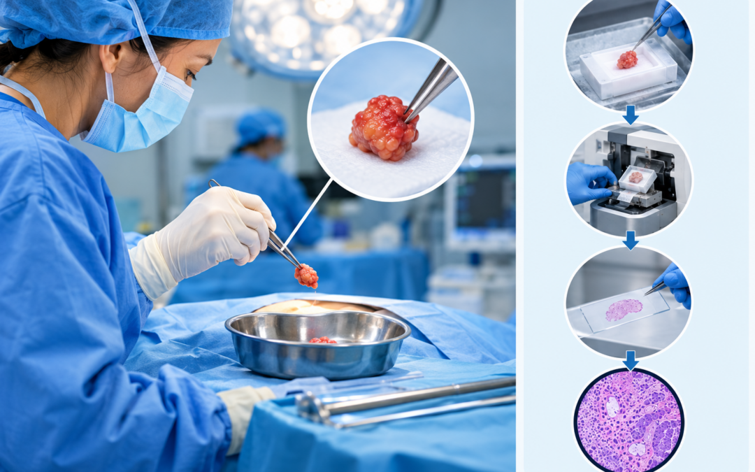
Three things decide it. First, hormonal blood and urine tests show whether the tumour is producing hormones, which points to what it is. Second, imaging reveals its size, density and shape, small, smooth and stable usually means benign, while large, irregular and dense raises concern. Third, where doubt remains, the tumour is removed and examined under a microscope for the final answer. Most turn out benign.

According to Dr. Sandeep Nayak, Surgical Oncologist in India, “The reassuring part is that most adrenal tumours are benign, so this isn’t usually a cancer hunt, it’s a careful sorting process. I look at the hormones first, then the imaging, the size, the density on CT, how contrast washes out, the shape of the edges. Those tell me a great deal before any surgery. When the picture is genuinely unclear, removing it and examining it gives the definitive answer. Guesswork has no place here.”

Found an adrenal mass and unsure what it means?

What Do Doctors Actually Check?

The assessment follows a clear sequence, and each step narrows down the answer.

  • Hormonal testing : Blood and urine tests show whether the tumour is functioning, pumping out hormones. This shapes both the diagnosis and how urgently it needs treating.
  • Size : Size is one of the strongest clues. Small tumours are usually benign, while the risk of cancer climbs as a tumour gets larger.
  • CT features : A CT scan looks at density and how contrast washes out. Benign adenomas are low density and wash out fast, cancers don’t.
  • The margins : Smooth, well defined edges point to benign. Irregular borders, patchy internal texture and invasion into nearby tissue suggest something more serious.

This structured workup is the foundation of proper adrenal tumor treatment, where the evaluation before surgery matters as much as the surgery itself.

Why Isn't a Biopsy the First Step?

For most tumours a needle biopsy is the go to, but the adrenal gland is a different case.

  • Biopsy is avoided : Sticking a needle into a suspected adrenal cancer risks spreading it, and it rarely helps. So doctors lean on imaging and hormones instead.
  • The pheo trap : If the tumour is a pheochromocytoma, a biopsy can trigger a dangerous blood pressure crisis. Hormonal testing rules this out first.
  • Surgery gives the answer : When a tumour looks suspicious, the safe route is removing it whole, then examining it. That confirms benign or malignant definitively.
  • Whole is better : Taking the tumour out intact, rather than sampling it, both diagnoses and treats it in one step, without risking any spread.

Understanding what these growths are in the first place, covered in our guide on adrenal tumors, makes this evaluation much clearer.

Why Choose Dr. Sandeep Nayak for Adrenal Tumor Care?

Dr. Sandeep Nayak is a surgical oncologist with 24 years behind him and a fellowship in laparoscopic and robotic onco-surgery. He treats the full range of adrenal tumours, from benign functioning adenomas and pheochromocytomas to adrenocortical carcinoma, using robotic and laparoscopic adrenalectomy. The approach starts with thorough biochemical and imaging assessment, since with adrenal tumours the evaluation before surgery decides everything, including whether an operation is even needed.

Adrenal tumours reward careful, methodical assessment more than most. Rushing to surgery on a benign adenoma helps no one, while missing the features of a cancer costs precious time. Reading the hormones and imaging correctly, knowing when to watch and when to operate, and preparing properly for tumours like pheochromocytoma, that judgement is what separates safe adrenal care from risky guesswork. The evaluation is where the real expertise lies.

Frequently Asked Questions

How do doctors know if an adrenal tumor is cancer?

Through hormonal testing, imaging features and, where doubt remains, surgery with histopathology.

Does size indicate cancer?

Larger adrenal tumours carry higher cancer risk, but size alone doesn’t confirm it.

What imaging features suggest cancer?

Irregular margins, large size, high CT density and slow contrast washout raise suspicion.

Is a biopsy used to diagnose it?

Rarely. Suspicious adrenal tumours are usually removed whole, then examined under a microscope.

References

  1. Differentiating benign from malignant adrenocortical tumors — National Library of Medicine
  2. Diagnostic workup of adrenal incidentaloma — National Library of Medicine

Disclaimer: This blog is for informational and educational purposes only and is not a substitute for professional medical advice or diagnosis.
Best Hospital for Robotic Lung Surgery?

Best Hospital for Robotic Lung Surgery?

The best hospital isn’t simply the one that owns a robot. Plenty of hospitals in Bangalore have the da Vinci system now. What separates them is the surgeon using it, how many robotic lung cases they actually do, whether there’s a real thoracic programme, and a team planning each case together. The machine doesn’t produce the outcome. The experienced hands guiding it do. That’s the distinction that matters.

According to Dr. Sandeep Nayak, Surgical Oncologist in India, “Patients see that a hospital has a robot and assume that settles it. It doesn’t. The da Vinci system is a tool, and what it produces depends entirely on who’s operating it. A surgeon who’s done robotic lung surgery hundreds of times is a different proposition from one whose hospital simply bought the machine. Ask about the surgeon’s volume, not the hospital’s equipment list. That’s where the real answer is.”

Looking for experienced robotic lung surgery in Bangalore?

What Makes a Hospital Good for This?

The right hospital for robotic lung surgery is defined by what surrounds the robot, not the robot itself.

  • The surgeon’s volume : This is the big one. A thoracic surgeon doing robotic lung cases regularly has judgement and skill that owning the machine can never supply.
  • A lung programme : A dedicated thoracic oncology setup, not the occasional lung case, means the whole team knows this surgery inside out.
  • The tumour board : Cases reviewed together by surgeons, oncologists and radiologists produce better plans than any single decision maker working alone.
  • Integrated recovery : Proper post-operative care, physiotherapy and follow up under one roof matters as much as the operation for a smooth result.

All of this sits within a serious robotic cancer surgery programme, where the platform is one part of a much bigger picture.

How Do You Judge It?

A few direct questions cut through the marketing and reveal what a hospital actually offers.

  • Ask the volume : How many robotic lung operations does the surgeon do each year? A specific, confident number tells you far more than a brochure.
  • Ask about the programme : Is there a dedicated thoracic team, or is lung surgery an occasional add on? The difference shows in outcomes.
  • Ask who decides : Is the case reviewed by a multidisciplinary board? Team based planning is a mark of a proper cancer centre.
  • Look past the hardware : Every hospital advertises its robot. The one worth choosing is where an experienced surgeon uses it at real volume.

This is the same logic covered in our guide on which hospital offers da Vinci robotic surgery, applied specifically to lung surgery.

Why Choose Dr. Sandeep Nayak for Robotic Lung Surgery?

Dr. Sandeep Nayak is a surgical oncologist with 24 years behind him and a fellowship in laparoscopic and robotic onco-surgery. He operates with the da Vinci system at associated hospitals in Bangalore, with over 15 years of robotic oncology experience and more than a thousand robotic cancer operations behind him, while consultation and planning stay centred at MACS Clinic. That combination of surgeon volume, a dedicated programme and team based planning is exactly what the criteria above describe.

Choosing where to have robotic lung surgery comes down to matching the setup to those standards, not to whichever hospital advertises its robot most. The platform is available in many places now, but the experience guiding it isn’t evenly spread. For a patient in Bangalore, the useful question isn’t which hospital has a da Vinci system, it’s which surgeon uses it for lung cancer at genuine volume, within a team, with the track record to show for it.

Frequently Asked Questions

What makes a hospital good for robotic lung surgery?

A high volume thoracic surgeon, a lung programme and a team, not just a robot.

Does having a robot make a hospital the best?

No. The surgeon’s experience with the robot matters far more than owning one.

Why does surgeon volume matter most?

A surgeon using the robot often builds the judgement that lowers complications and improves outcomes.

What else should a good hospital have?

A thoracic programme, tumour board planning and integrated post-operative recovery care.

References

  1. Hospital volume and outcomes in robotic lung resection — National Library of Medicine
  2. Surgeon experience and robotic thoracic surgery outcomes — National Library of Medicine

Disclaimer: This blog is for informational and educational purposes only and is not a substitute for professional medical advice or diagnosis.

What Is Laparoscopic Adrenalectomy Recovery Like?

What Is Laparoscopic Adrenalectomy Recovery Like?

Recovery after laparoscopic adrenalectomy is quick compared to open surgery. Most patients stay in hospital just one to three days, walk the same or next day, and manage pain well with oral tablets. Light activity comes back within a week or two, with fuller normal activity by around a month. If the tumour was hormone producing, blood tests continue afterward while the body readjusts. Overall, it’s a gentle recovery for a major gland’s removal.

According to Dr. Sandeep Nayak, Surgical Oncologist in India, “What surprises most patients is how quickly they’re back on their feet. We have people walking the day of surgery and home within a couple of days. The small incisions mean far less pain than the open operation used to involve. The one thing I do watch closely is hormone levels afterward, especially if the tumour was producing excess hormone, because the body needs a little time to find its new balance.”

Preparing for adrenal surgery and wondering about recovery?

What Happens in the First Few Days?

The early recovery period focuses on getting the patient up, comfortable and eating again.

  • Hospital stay : Most patients go home within one to three days, a fraction of what open adrenal surgery once required.
  • Early walking : Getting up and moving the same day or the next is encouraged. It helps circulation and speeds the whole recovery along.
  • Pain control : Discomfort from the small incisions is usually mild to moderate, managed well with oral pain medication rather than anything stronger.
  • Return to eating : Diet progresses quickly from liquids back to normal food, usually within the first day or two after surgery.

This gentle early course is one of the real advantages built into modern adrenal tumor treatment, where the minimally invasive approach shortens the whole journey.

What Happens Over the Following Weeks?

The weeks after discharge bring a steady, predictable return toward normal life.

  • First two weeks : Light activity, short walks and normal daily tasks return fairly quickly. Heavy lifting and strenuous exercise still wait a little longer.
  • Around a month : Most patients are back to their usual routine, including work for many, by roughly four weeks after surgery.
  • Hormone monitoring : If the tumour was producing excess hormone, blood tests track levels afterward, since the body needs time to rebalance and sometimes needs temporary support.
  • Wound healing : The small incisions heal quickly and with minimal scarring, one of the clearer visible benefits of the laparoscopic approach.

Getting to this recovery safely starts with the surgery itself, part of the broader picture of adrenal tumor surgery and how these tumours are managed.

Why Choose Dr. Sandeep Nayak for Adrenal Surgery?

Dr. Sandeep Nayak is a surgical oncologist with 24 years behind him and a fellowship in laparoscopic and robotic onco-surgery. He performs laparoscopic and robotic adrenalectomy with the technique and hormone awareness this surgery demands, since a smooth recovery depends on more than the operation alone. The approach includes planning for hormone monitoring after surgery, particularly for functioning tumours, so the recovery is followed through properly rather than ending at discharge.

A well done laparoscopic adrenalectomy gives patients back their normal life remarkably fast for major gland surgery. But the recovery isn’t only about the incisions healing, it’s about the body’s hormones settling into their new normal, especially after a functioning tumour is removed. Following that through with proper monitoring, alongside a technically clean operation, is what turns a good surgery into a genuinely smooth recovery.

Frequently Asked Questions

How long is the hospital stay after laparoscopic adrenalectomy?

Usually one to three days, much shorter than open adrenal surgery.

When can normal activity resume?

Light activity within one to two weeks, fuller activity by around a month.

Is pain significant after this surgery?

Pain is usually mild to moderate and well controlled with oral medication.

Does hormone monitoring continue after surgery?

Yes, especially after a hormone producing tumour, since levels need time to normalise.

References

  1. Recovery outcomes after laparoscopic adrenalectomy — National Library of Medicine
  2. Laparoscopic versus open adrenalectomy comparison — National Library of Medicine

Disclaimer: This blog is for informational and educational purposes only and is not a substitute for professional medical advice or diagnosis.

What Is Robotic Lung Cancer Surgery?

What Is Robotic Lung Cancer Surgery?

Robotic lung cancer surgery removes a lung tumour through a few small chest incisions, using a robotic system the surgeon controls. There’s no large cut, no spreading of the ribs. The surgeon sits at a console with a magnified 3D view and instruments that move with more precision than the human wrist. It’s used mainly for early stage lung cancer, and the recovery is far gentler than open surgery.

According to Dr. Sandeep Nayak, Surgical Oncologist in India, “With robotic lung surgery, I’m operating through keyhole incisions with a view and steadiness that open surgery can’t match. The 3D magnification lets me see the anatomy beautifully, and the wristed instruments reach places that are awkward otherwise. For the patient, that means no big chest incision, less pain and a quicker recovery. The cancer clearance is every bit as thorough. That combination is why it’s become so valuable.”

Considering minimally invasive surgery for lung cancer?

How Does Robotic Lung Surgery Work?

The technology changes how the surgeon operates, but the goal, removing the cancer completely, stays the same.

  • Small incisions : A few keyhole cuts replace the large chest opening of traditional surgery. No rib spreading, which is a major source of pain and slow recovery.
  • The console : The surgeon operates from a console, controlling robotic arms in real time. Every movement is the surgeon’s, scaled and steadied by the system.
  • 3D vision : A magnified, three dimensional view of the chest gives far better depth and detail than the naked eye, helping precise, safe dissection.
  • Wristed instruments : The instruments bend and rotate like a human wrist, only more, reaching tight spaces in the chest that rigid tools struggle with.

This precision is the heart of modern robotic cancer surgery, applied here to the delicate, confined space of the chest.

What Are the Benefits and Who Is It For?

The advantages are real, and they matter most for the right patient.

  • Faster recovery : Smaller incisions and no rib spreading mean less pain and a quicker return home. Many patients are up and moving very soon after.
  • Same cancer control : This is the key point. Survival, clear margins and lymph node clearance match open surgery. Nothing is traded for the gentler approach.
  • Precise node clearance : The 3D view and fine instruments make thorough lymph node removal easier, which is essential for accurate staging and control.
  • Best for early stage : It suits early stage lung cancer especially, and selected more advanced cases. A team assessment decides who’s a good candidate.

It sits alongside VATS surgery as the two leading minimally invasive options, each excellent in the right hands.

Why Choose Dr. Sandeep Nayak for Robotic Lung Surgery?

Dr. Sandeep Nayak is a surgical oncologist with 24 years behind him and a fellowship in laparoscopic and robotic onco-surgery. He performs both robotic and VATS lung surgery, choosing the approach that fits the individual tumour and patient rather than favouring one by default. With over 15 years of robotic oncology experience, he brings the volume and judgement that minimally invasive lung surgery genuinely demands. That experience is what makes the technique safe and effective.

The value in robotic lung surgery isn’t the machine itself, it’s the surgeon guiding it. The platform offers precision and vision, but only experienced hands turn that into complete, safe cancer removal with a smooth recovery. For a patient with early stage lung cancer, robotic surgery in a high volume centre offers a real chance at cure with far less of the trauma that open chest surgery once meant. Matching the method to the case is the whole craft.

Frequently Asked Questions

What is robotic lung cancer surgery?

Removing a lung tumour through small chest incisions using a robotic surgical system.

How is it different from open surgery?

It uses small incisions instead of opening the chest, so recovery is faster.

Who is suitable for robotic lung surgery?

Mainly early stage lung cancer patients fit for minimally invasive surgery.

Is it as effective as open surgery?

Yes. Survival, margins and lymph node clearance match open surgery in suitable cases.

References

  1. Robotic versus video-assisted thoracoscopic lobectomy meta-analysis — National Library of Medicine
  2. Pain and recovery after robotic lobectomy for lung cancer — National Library of Medicine

Disclaimer: This blog is for informational and educational purposes only and is not a substitute for professional medical advice or diagnosis.

Can Lung Cancer Be Detected on a Chest X-Ray?

Can Lung Cancer Be Detected on a Chest X-Ray?

A chest X-ray can show lung cancer, but it misses a lot. It picks up larger tumours reasonably well. Small, early ones often slip past it entirely. By the time a cancer is clearly visible on an X-ray, it’s frequently already advanced. A normal X-ray doesn’t mean there’s no cancer. That’s the crucial catch. For proper early detection, a low dose CT scan is far more reliable.

According to Dr. Sandeep Nayak, Surgical Oncologist in India, “A chest X-ray is useful, but I never want a patient to think a clear one is a guarantee. Small tumours hide easily, behind the heart, behind a rib. Most lung cancers that get missed are missed on X-rays, not CT. If someone is genuinely at risk, a low dose CT finds things an X-ray simply can’t. The X-ray has its place, but it isn’t the final word on lung cancer.”

Have a symptom or risk that needs proper checking?

What Can a Chest X-Ray Show?

An X-ray is a useful first look, but it’s important to know its reach and its blind spots.

  • Larger masses : A chest X-ray shows bigger tumours reasonably well. If a mass is large enough, it usually appears as a visible shadow.
  • A first step : It’s quick, cheap and widely available, which makes it a common first test when someone has chest symptoms.
  • Other clues : It can reveal related signs, like fluid around the lung or a collapsed segment, that prompt further investigation.
  • The small ones slip : Its real weakness is small tumours. A cancer in its early, most treatable stage often doesn’t show up at all.

Because of these limits, proper lung cancer treatment planning relies on CT and other imaging rather than an X-ray alone.

Why Isn't It Enough on Its Own?

The gap between what an X-ray shows and what a CT shows is what matters most here.

  • Misses early tumours : This is the big one. The majority of missed lung cancers are missed on chest X-rays, precisely when catching them counts most.
  • Hidden spots : Tumours behind the heart, ribs or diaphragm can hide from an X-ray entirely, sitting in blind spots the flat image can’t separate.
  • CT is sharper : A low dose CT builds detailed cross sections, spotting nodules far too small for an X-ray. For screening, it’s the proven tool.
  • Clear isn’t cleared : A normal X-ray is reassuring but not conclusive. Anyone with persistent symptoms or real risk deserves a closer look regardless.

For those at highest risk, understanding smoking and lung cancer explains who should consider CT screening rather than relying on an X-ray.

Why Choose Dr. Sandeep Nayak for Lung Cancer Care?

Dr. Sandeep Nayak is a surgical oncologist with 24 years behind him and a fellowship in laparoscopic and robotic onco-surgery. He treats lung cancer with VATS and robotic thoracic surgery, and he’s clear with patients that the right imaging is what makes early, curable diagnosis possible. The approach means not stopping at a normal X-ray when symptoms or risk suggest otherwise, since the whole outcome can turn on getting the right scan at the right time.

The imaging choice shapes everything downstream. A lung cancer found early on CT, while it’s small and operable, is a completely different situation from one found late on an X-ray after it’s grown. Knowing when an X-ray is enough and when it isn’t, and moving to CT without delay for those at risk, is the judgement that turns detection into a real chance at cure.

Frequently Asked Questions

Can lung cancer be detected on a chest X-ray?

It can show larger tumours, but often misses small, early lung cancers.

Does a normal chest X-ray rule out lung cancer?

No. A normal X-ray doesn’t rule out cancer, since small tumours can be missed.

What is better than an X-ray for detection?

Low dose CT is far more sensitive and detects much smaller lung tumours.

Why are tumours missed on X-ray?

Small tumours, or those hidden behind the heart, ribs or diaphragm, can be missed.

References

  1. Low dose CT versus chest radiography in lung cancer screening — National Library of Medicine
  2. Early detection of lung cancer in high-risk patients — ClinicalTrials.gov

Disclaimer: This blog is for informational and educational purposes only and is not a substitute for professional medical advice or diagnosis.

Call Now Button