support@macsclinic.com
+91 9482202240
 Does an IHC Test Mean Cancer?

 Does an IHC Test Mean Cancer?

The Immunohistochemistry (IHC) test does not necessarily mean that you have cancer, but it is a specialized diagnostic procedure that is often used to detect, classify, and stage cancer cells in the body. This procedure is used to analyze tissues in the body to detect certain proteins to ascertain if the tumor is cancerous or non-cancerous.

According to Dr. Sandeep Nayak, cancer specialist in Bangalore, “An IHC test doesn’t confirm cancer by itself but it tells us things about cells that completely change how we treat whatever we’ve already found.”

According to Dr. Sandeep Nayak, cancer specialist in Bangalore, “A CT scan shows us a picture of what’s there but a picture is not a diagnosis and confusing the two is where patients and sometimes doctors go wrong.”

What Is an IHC Test Actually Doing to Your Tissue Sample?

Most patients who’ve been told an IHC has been ordered have absolutely no idea what that means in practice. Here’s what’s genuinely happening in that laboratory with your sample.

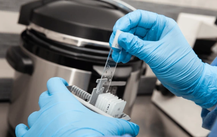
  • It Uses Antibodies to Tag Specific Proteins Inside Your Cells: IHC works by applying specially designed antibodies to tissue sections that bind to particular proteins and then stain them visible colours under a microscope showing the pathologist exactly what’s present at a molecular level.
  • It Tells Pathologists Where a Cancer Started When the Origin Isn’t Obvious: When cancer is found in lymph nodes or a distant organ without a clear primary source IHC markers help identify whether those cells originally came from the breast, lung, colon or another organ entirely.
  • It Identifies Whether a Cancer Will Respond to Specific Treatments: HER2 status in breast cancer, PD-L1 expression for immunotherapy eligibility and hormone receptor status are all determined through IHC and these results directly decide which cancer treatment pathway you go down.
  • It Distinguishes Between Cancer Types That Look Similar Under Basic Microscopy: Two tumours can look almost identical on a standard biopsy but behave completely differently and need completely different treatment and IHC separates them by their molecular fingerprint rather than just their appearance.

The newly developed laparoscopic surgery  techniques can facilitate the achievement of effective removal of the tumour in smaller incisions and less time of recovery in the right patients in the event of early diagnosis and localisation of the cancer.

What Does an IHC Result Actually Tell Your Doctor About Your Specific Case?

This is the part that matters most to you sitting with that report. Not the science. What it means for your treatment. What happens next. Here’s how IHC results change real clinical decisions.

  • Hormone Receptor Status Decides Whether Hormone Therapy Will Work for You: ER and PR positive results in breast cancer mean hormone blocking therapy is likely to be highly effective while negative results shift treatment toward chemotherapy and targeted drugs instead.
  • HER2 Status Opens or Closes the Door to Targeted Therapy: HER2 positive cancer responds to trastuzumab and other targeted agents that can dramatically improve outcomes but without IHC confirmation of HER2 status that entire treatment pathway simply doesn’t get considered.
  • Ki-67 Index Tells Your Oncologist How Fast the Cancer Is Growing: This IHC marker measures the proportion of actively dividing cells giving a direct indication of tumour aggressiveness that influences how urgently and how intensively cancer treatment needs to begin.
  • Lymphoma Subtyping Through IHC Completely Changes the Treatment Protocol: Hodgkin lymphoma and various non-Hodgkin subtypes look similar under basic microscopy but respond to entirely different chemotherapy regimens and IHC is what separates them accurately and reliably.

In cases of cancers where a high degree of accuracy in tumour removal is demanded in anatomically complex regions, innovative robotic surgery technologies are becoming a popular method of enhancing the accuracy of surgery and recovery in patients..

Why Choose Dr. Sandeep Nayak for Cancer Treatment in Bangalore?

Dr. Sandeep Nayak has spent over 24 years building cancer treatment plans on the kind of molecular precision that IHC testing makes possible. He doesn’t treat cancer types generically. He treats the specific subtype that the IHC has identified in your specific tissue with the specific treatment approach that subtype actually responds to. As one of the most trusted cancer specialists in Bangalore he works with detailed pathology reports the way a good detective works with evidence. Every marker. Every staining pattern. Every molecular clue. All of it feeds into a treatment plan that’s built entirely around your cancer’s actual biology rather than what it looked like it might be before the detailed testing was done.

Frequently Asked Questions

Does being sent for an IHC test mean your doctor already knows you have cancer?

Not necessarily. IHC is ordered whenever tissue examination needs molecular level detail which includes confirming cancer type, ruling out malignancy or identifying cancer origin in unclear cases.

How long does an IHC test result typically take to come back?

IHC results generally take five to ten working days depending on the laboratory and the number of markers being tested on your specific tissue sample.

Can an IHC test result change what type of cancer treatment you receive?

 Absolutely yes. IHC results directly determine hormone therapy eligibility, targeted therapy suitability, chemotherapy protocol selection and overall cancer treatment pathway for most solid tumours.

Is IHC testing available at all cancer centres in India or only specialised ones?

IHC testing requires specialised equipment and trained pathologists and is most reliably performed at dedicated cancer centres and academic hospitals rather than general diagnostic laboratories.

Reference links:

Can a CT Scan Detect Cancer?

Can a CT Scan Detect Cancer?

Yes. But not always. And not definitively on its own. That second part is the bit nobody explains properly and it’s the part that actually matters most when you’re sitting with a CT report in your hands trying to figure out what it means for your life. A CT scan is powerful. Really powerful. But it has limits. Real ones. And understanding those limits is just as important as understanding what the scan can do.

According to Dr. Sandeep Nayak, cancer specialist in Bangalore, “A CT scan shows us a picture of what’s there but a picture is not a diagnosis and confusing the two is where patients and sometimes doctors go wrong.”

What Can a CT Scan Actually Find?

People walk in thinking a CT scan is either going to find cancer or rule it out. Clean and simple. But it’s genuinely more complicated than that and you deserve to understand how.

  • It Finds Masses and Tumours That Nothing Else Would Catch at That Stage: A contrast enhanced CT can pick up tumours just a few millimetres across in the liver, lungs, kidneys, pancreas and lymph nodes that would be completely invisible to any physical examination by any doctor.
  • It Shows Whether Cancer Has Moved Beyond Where It Started: Chest abdomen and pelvis CT together gives oncologists a real map of whether cancer has crept into lymph nodes, nearby organs or distant sites and that map is what makes accurate staging and cancer treatment planning actually possible.
  • It Guides the Needle Exactly Where It Needs to Go for Biopsy: When something suspicious shows up CT guided biopsy puts a needle into abnormal tissue with pinpoint accuracy so pathology can finally tell you whether what’s there is actually cancer or something far less serious.
  • It Watches What Treatment Is Doing to the Tumour Over Time: Once cancer treatment starts repeat CT scans at regular intervals show whether tumours are shrinking, staying the same or growing giving your oncologist real evidence to either continue the plan or change it.

In cases of cancers where a high degree of accuracy in tumour removal is demanded in anatomically complex regions, innovative robotic surgery technologies are becoming a popular method of enhancing the accuracy of surgery and recovery in patients.

What Can a CT Scan Actually Miss? Because This Part Matters.

Nobody talks about this clearly enough. And patients who don’t know it make decisions based on false reassurance that can cost them months they don’t have.

  • A Normal CT Does Not Mean Cancer Has Been Completely Ruled Out: Early blood cancers, tiny tumours below detection thresholds and cancers that spread along surfaces rather than forming distinct masses can all be actively growing while your CT appears completely unremarkable.
  • Finding a Mass on CT Is Not the Same Thing as a Cancer Diagnosis: The scan shows something is there. Pathology from a biopsy tells you what it actually is. These are two separate steps and skipping the second one because the first one looked worrying is never acceptable clinical practice.
  • Certain Body Parts Are Simply Better Seen on MRI Than CT: Brain tumours, spinal cord disease, soft tissue masses and specific pelvic cancers show up with far more clarity on MRI and choosing the wrong scan for the wrong body part genuinely delays diagnosis in ways that matter.
  • Radiation Exposure From Repeated CT Scans Is Something Worth Thinking About: A single CT scan is considered safe. But repeated scans every few months over years involves cumulative radiation exposure that deserves thoughtful clinical justification rather than being ordered just to reassure someone who’s anxious.

The newly developed laparoscopic surgery  techniques can facilitate the achievement of effective removal of the tumour in smaller incisions and less time of recovery in the right patients in the event of early diagnosis and localisation of the cancer.

Why Choose Dr. Sandeep Nayak for Cancer Treatment in Bangalore?

Dr. Sandeep Nayak has spent over 24 years doing something that sounds simple but is genuinely rare. He looks at CT scans in the context of everything else he knows about the person sitting across from him. Their symptoms. Their history. Their markers. Their story. As one of the most trusted cancer specialists in Bangalore he doesn’t stop investigating when a scan looks normal and symptoms keep pointing somewhere. He keeps asking. He keeps testing. He keeps looking. Because in his experience the patients who needed that persistence most were precisely the ones whose early imaging appeared deceptively reassuring to everyone who looked at it before him.

Frequently Asked Questions

Can cancer genuinely be present in your body while a CT scan shows nothing?

Yes, very small tumours, early blood cancers and certain cancer types that spread along surfaces rather than forming masses can be active while CT appears completely normal.

Does getting a CT scan with contrast actually make a meaningful difference to detection?

Yes, contrast enhancement significantly improves tumour visibility by highlighting blood flow differences between cancerous and normal tissue making previously invisible masses much clearer.

How long before you actually get results after a CT scan is done?

Radiologist reports typically come back within 24 to 72 hours but what those findings actually mean for your situation needs to be interpreted with your oncologist in proper clinical context.

Is a PET scan genuinely better than CT for finding cancer that has spread?

PET scans detect metabolically active cancer cells throughout the entire body and are often more sensitive for spread than CT alone making them powerful complementary investigations used together.

Reference links:

Cancers That Cause Fluid in the Abdomen

Cancers That Cause Fluid in the Abdomen

Fluid in the abdominal cavity, also known as malignant ascites, is often a result of cancers that metastasize to the peritoneum, which includes ovarian, pancreatic, liver, stomach, and colorectal cancers. This results from metastatic involvement of the peritoneum that leads to leakage or lymphatic obstruction.

According to Dr. Sandeep Nayak, cancer specialist in Bangalore, “Malignant ascites tells us the cancer has reached the peritoneum and that finding changes the cancer treatment conversation significantly in ways patients need to understand clearly.”

Which Cancers Most Commonly Cause Fluid to Build Up in the Abdomen?

Most people with abdominal swelling assume it’s a digestive problem. A liver issue. Something they ate. And sometimes they’re right. But when the fluid keeps coming back or arrives without an obvious explanation these are the cancers your doctor is thinking about.

  • Ovarian Cancer Is the Single Most Common Cause of Malignant Ascites: Ovarian cancer spreads along the peritoneal lining with a particular efficiency that causes fluid to accumulate in large volumes and abdominal bloating that keeps returning despite drainage is one of its most consistent presentations in women.
  • Colorectal Cancer Spreads to the Peritoneum and Drives Fluid Accumulation: When colorectal cancer reaches the peritoneal cavity it seeds cancer cells across the abdominal lining and those cells trigger inflammatory fluid production that accumulates progressively and doesn’t resolve on its own.
  • Stomach Cancer Causes Peritoneal Spread That Produces Significant Ascites: Gastric cancer has a particular tendency to seed the peritoneum early and ascites appearing alongside upper abdominal discomfort and unexplained weight loss in someone over 50 is a combination that needs urgent investigation.
  • Liver Cancer and Secondary Liver Metastases Both Cause Fluid Through Different Mechanisms: Primary liver cancer disrupts liver function directly while metastatic cancer in the liver causes portal hypertension and peritoneal irritation both of which independently drive fluid accumulation in the abdominal cavity.

The newly developed laparoscopic surgery  techniques can facilitate the achievement of effective removal of the tumour in smaller incisions and less time of recovery in the right patients in the event of early diagnosis and localisation of the cancer.

What Does Malignant Ascites Actually Mean for Your Cancer Treatment Options?

This is the part nobody sits down and explains properly. And patients deserve to understand it because it changes what happens next in very real and specific ways.

  • It Usually Means Cancer Has Reached the Peritoneal Cavity: The peritoneum is the membrane lining your entire abdominal cavity and when cancer seeds itself there it creates a situation that’s more complex to treat than a single contained tumour in one organ.
  • Drainage Relieves Symptoms But Doesn’t Treat the Underlying Cancer: Paracentesis drains the fluid and makes you feel better within hours but the fluid comes straight back unless the cancer causing it is being actively treated because the source of the problem is still there producing more.
  • HIPEC Surgery Offers a Genuinely Curative Option for Selected Patients: Hyperthermic intraperitoneal chemotherapy delivered directly into the abdominal cavity during surgery can achieve complete peritoneal clearance in carefully selected patients with certain cancer types and this treatment has changed outcomes dramatically for eligible cases.
  • The Earlier Peritoneal Spread Is Found the More Treatment Options Remain Open: Patients with limited peritoneal disease detected early have access to surgical and chemotherapy options that simply aren’t available once fluid accumulation becomes massive and widespread throughout the entire abdominal cavity.

In cases of cancers where a high degree of accuracy in tumour removal is demanded in anatomically complex regions, innovative robotic surgery technologies are becoming a popular method of enhancing the accuracy of surgery and recovery in patients..

Why Choose Dr. Sandeep Nayak for Cancer Treatment in Bangalore?

Dr. Sandeep Nayak has spent over 24 years treating peritoneal cancers and malignant ascites from ovarian, colorectal, gastric and liver primaries using cytoreductive surgery and HIPEC procedures that offer genuinely curative outcomes for carefully selected patients. As one of the most experienced cancer specialists in Bangalore performing these complex procedures he doesn’t just drain the fluid and send patients home. He investigates the source. He stages the peritoneal disease properly. He identifies whether HIPEC candidacy exists. And he builds a cancer treatment plan that addresses what’s actually driving the ascites not just its most visible symptom. Because fluid in the abdomen is never the real problem. It’s always the sign of one.

Frequently Asked Questions

Can abdominal fluid from cancer be permanently resolved with treatment?

 Yes, in selected patients with limited peritoneal spread cytoreductive surgery combined with HIPEC can achieve complete peritoneal clearance and prevent ascites from returning permanently.

How do doctors confirm whether abdominal fluid is caused by cancer?

 Ascitic fluid analysis through paracentesis combined with CT scan PET imaging and tumour marker blood tests together confirm whether ascites has a malignant origin.

Which cancer type most commonly causes massive abdominal fluid accumulation?

Ovarian cancer produces the largest volumes of malignant ascites most consistently and is the first cancer investigated when a woman presents with unexplained abdominal fluid.

Can ascites caused by cancer be drained repeatedly to manage symptoms?

Yes, repeated paracentesis manages symptoms effectively but fluid returns quickly without active cancer treatment making systemic therapy or surgical intervention essential for lasting control.

Reference links:

Can a DEXA Scan Show Cancer?

Can a DEXA Scan Show Cancer?

Not directly. A DEXA scan was designed to measure bone density. That’s its job. That’s what it was built for. But here’s what most people sitting with an unexpected DEXA result don’t know. Sometimes while doing that job it picks up changes in bone structure that raise questions worth investigating properly. And occasionally those questions lead somewhere more serious than osteoporosis.

According to Dr. Sandeep Nayak, cancer specialist in Bangalore,
“A DEXA scan isn’t a cancer screening tool but sometimes it finds things in bones that point toward something we absolutely need to investigate further.”

What Does a DEXA Scan Actually Do and What Can It Pick Up?

Most people getting a DEXA scan are thinking about bone loss. Osteoporosis. Fracture risk. Nobody walks into that appointment expecting a cancer conversation. But sometimes that’s exactly where it goes.

  • It Measures Bone Mineral Density With Remarkable Precision: DEXA uses low dose X-ray beams to measure how dense and strong your bones are at the spine and hip giving doctors a precise picture of bone health that no other scan provides as accurately.
  • It Can Detect Unusual Bone Changes That Weren’t Expected: When bone density appears abnormally low in a pattern that doesn’t match typical osteoporosis or when focal bone lesions show up in the scan field something more than age related bone loss may be happening inside those bones.
  • Cancers That Spread to Bone Sometimes Change Bone Density in Ways DEXA Picks Up: Bone metastases from breast, prostate, lung, kidney and thyroid cancers alter bone structure and density in ways that occasionally appear on DEXA scans before the patient has any pain or obvious symptoms at all.
  • Multiple Myeloma Is the Blood Cancer Most Closely Linked to Unexpected DEXA Findings: This bone marrow cancer actively destroys bone from the inside and significantly abnormal DEXA results in someone without clear osteoporosis risk factors should prompt investigation for myeloma specifically.

In cases of cancers where a high degree of accuracy in tumour removal is demanded in anatomically complex regions, innovative robotic surgery technologies are becoming a popular method of enhancing the accuracy of surgery and recovery in patients.

When Should a DEXA Result Actually Prompt Cancer Investigation?

This is the part most patients never get told clearly. And it matters because the difference between acting on a finding and filing it away is sometimes the difference between catching something early and missing the window entirely.

  • Severe Unexplained Bone Loss in Someone Without Osteoporosis Risk Factors: If you’re relatively young, haven’t had long term steroid use and don’t have other obvious causes for bone loss but your DEXA shows severely low density that pattern needs a full blood workup not just a bone density recheck.
  • Focal Lesions or Asymmetric Changes That Appear in the Scan Field: DEXA occasionally captures part of the spine or pelvis where focal abnormalities appear and any localised bone lesion seen incidentally needs follow up imaging with CT or MRI to understand what it actually represents.
  • Rapidly Worsening Bone Density Between Two Scans Done Close Together: Normal bone loss happens slowly over years. Density that drops dramatically in twelve to eighteen months is losing bone far faster than biology normally allows and that speed demands an explanation beyond just ageing.
  • DEXA Findings Alongside Symptoms That Don’t Fit a Simple Osteoporosis Story: Bone pain that wakes you at night, unexplained fatigue, weight loss or anaemia sitting alongside a concerning DEXA result is a combination that needs oncology eyes on it without delay.

The newly developed laparoscopic surgery  techniques can facilitate the achievement of effective removal of the tumour in smaller incisions and less time of recovery in the right patients in the event of early diagnosis and localisation of the cancer.

Why Choose Dr. Sandeep Nayak for Cancer Treatment in Bangalore?

Dr. Sandeep Nayak has spent over 24 years treating cancers that don’t always introduce themselves in obvious ways. Bone metastases from thyroid, colorectal and gastrointestinal cancers. Multiple myeloma presentations that started as inexplicable bone density changes. Complex cases where the first clue was hiding inside a scan result that most people would never connect to oncology at all. As one of the most trusted cancer specialists in Bangalore he reads unexpected findings with genuine curiosity rather than dismissal. He investigates what doesn’t fit. He asks the questions that turn a confusing result into a clear diagnosis. And he builds a cancer treatment plan around what he actually finds rather than what was expected going in.

Frequently Asked Questions

Does a normal DEXA scan completely rule out bone cancer or bone metastases?

 No, DEXA is not a cancer screening tool and normal bone density doesn’t exclude bone metastases or early myeloma which need dedicated imaging and blood tests to properly evaluate.

Which cancers most commonly spread to bone and affect DEXA scan results?

 Breast, prostate, lung, kidney and thyroid cancers most commonly spread to bone while multiple myeloma directly destroys bone from within the marrow affecting density measurements significantly.

What tests should follow an unexplained abnormal DEXA scan result?

Full blood count, serum protein electrophoresis, PSA, tumour markers and follow up MRI or CT of the abnormal area together give the most complete diagnostic picture.

Can bone density improve again after cancer treatment for bone metastases?

 Yes, some cancer treatments including bisphosphonates and denosumab alongside primary cancer treatment can stabilise and in some cases partially restore bone density affected by metastatic disease.

Reference links:

 Does High MCV Mean Cancer?

 Does High MCV Mean Cancer?

Not automatically. But it’s worth understanding what high MCV actually means before you either panic or dismiss it completely. MCV stands for mean corpuscular volume. It measures the size of your red blood cells. When they’re bigger than they should be something in your body is off. Usually something fixable. But occasionally something that deserves a much closer look than a repeat blood test in three months.

According to Dr. Sandeep Nayak, cancer specialist in Bangalore, “High MCV on its own rarely means cancer but when it keeps coming back elevated without a clear explanation it’s a signal worth taking seriously with proper investigation.”

What Does High MCV Actually Mean in Real Terms?

People see this flagged on their blood report and either Google themselves into a spiral or completely ignore it. Neither helps. Here’s what’s genuinely happening when that number comes back high.

  • Normal MCV Sits Between 80 and 100 Femtolitres in Most Adults: Anything above 100 fL is called macrocytosis meaning your red blood cells are larger than they should be and while this sounds alarming it has many causes most of which aren’t cancer at all.
  • Vitamin B12 and Folate Deficiency Are the Most Common Culprits by Far: These two nutritional deficiencies prevent red blood cells from dividing properly making them grow abnormally large and this is the explanation behind the majority of high MCV results seen in routine blood work across India.
  • Alcohol Use Raises MCV Consistently and Often Gets Overlooked: Heavy regular alcohol consumption directly affects how bone marrow produces red blood cells and elevated MCV in someone who drinks regularly without B12 deficiency is often simply the body’s response to chronic alcohol exposure.
  • But When the Common Causes Are Ruled Out the Conversation Changes: An MCV that stays elevated after B12 and folate are corrected, without alcohol as an explanation, without thyroid disease or liver disease, is the result that genuinely warrants cancer investigation without further delay.

In cases of cancers where a high degree of accuracy in tumour removal is demanded in anatomically complex regions, innovative robotic surgery technologies are becoming a popular method of enhancing the accuracy of surgery and recovery in patients.

Which Cancers Are Most Commonly Linked to High MCV?

Not every cancer drives MCV up. But specific ones do it consistently enough that haematologists actively look for them when common causes have been excluded properly.

  • Myelodysplastic Syndrome Is the Most Direct Association: MDS is a bone marrow disorder that often sits on the borderline between precancer and cancer and high MCV alongside anaemia and low blood counts is one of its most consistent early presentations in adults over 60.
  • Acute Myeloid Leukaemia Can Present With Elevated MCV Alongside Other Blood Count Changes: AML disrupts normal bone marrow function dramatically and abnormal red cell size alongside falling white cells and platelets is a pattern that needs same week haematology review not watchful waiting.
  • Certain Solid Tumours Cause High MCV Through Paraneoplastic Effects: Some cancers including liver cancer and certain lung cancers affect bone marrow function indirectly through hormonal and inflammatory signals producing macrocytosis without direct bone marrow invasion being present.
  • Chemotherapy Treatment for Any Cancer Frequently Causes MCV to Rise: If you’ve had cancer treatment recently high MCV is often a direct side effect of chemotherapy drugs affecting how your bone marrow produces red blood cells rather than a sign of new cancer activity.

The newly developed laparoscopic surgery  techniques can facilitate the achievement of effective removal of the tumour in smaller incisions and less time of recovery in the right patients in the event of early diagnosis and localisation of the cancer.

Why Choose Dr. Sandeep Nayak for Cancer Treatment in Bangalore?

Dr. Sandeep Nayak has spent over 24 years treating blood cancers and haematological conditions that often start as unexplained abnormalities on routine blood reports that nobody quite connected to anything serious soon enough. As one of the most trusted cancer specialists in Bangalore he doesn’t stop at the obvious explanation when the obvious explanation doesn’t actually fit. He looks at the full blood picture. The trends over time. The symptoms alongside the numbers. The clinical context that turns a flagged result into a real answer. His patients consistently describe someone who found what others missed. Not because he’s looking harder necessarily. But because he keeps looking after everyone else has stopped.

Frequently Asked Questions

Can high MCV go back to normal without cancer treatment if it's nutritional?

 Yes, MCV caused by B12 or folate deficiency normalises within weeks to months after appropriate supplementation and addressing the underlying nutritional deficiency properly.

What blood tests should follow a persistently high MCV with normal B12 levels?

Full blood count with differential, reticulocyte count, liver function tests, thyroid function and bone marrow biopsy together give the most complete picture of what’s driving persistent macrocytosis.

How quickly should you act on a persistently high MCV that keeps coming back?

 If MCV stays elevated across three or more consecutive blood tests despite correcting B12 and folate you should see a specialist within weeks not months for proper investigation.

Can children get high MCV linked to cancer or is it mainly an adult finding?

High MCV linked to haematological malignancy occurs predominantly in adults particularly over 60 though childhood leukaemias can also produce abnormal blood count patterns including MCV changes.

Reference links:

What Is ESR Level in Cancer Patients?

What Is ESR Level in Cancer Patients?

There’s no fixed number. That’s the truth nobody wants to hear but it’s the only honest starting point. ESR or erythrocyte sedimentation rate measures how fast your red blood cells settle in a test tube. When something is inflaming your body those cells clump and sink faster. Cancer does that. But so does infection. So does arthritis. So does pregnancy. The number alone never tells the whole story.

According to Dr. Sandeep Nayak, cancer specialist in Bangalore, “ESR is one of those results that tells you your body is reacting to something but figuring out exactly what that something is requires a lot more than just looking at one number.”

What Do Elevated ESR Levels Actually Mean in Real Terms?

Most people get this result handed to them with very little explanation. They go home and Google it at midnight. And what they find either terrifies them or confuses them completely. Here’s what’s genuinely happening.

  • Normal ESR Sits Below 20 mm/hr for Men and Below 30 mm/hr for Women Generally: These ranges shift with age so an older adult naturally has a higher baseline and what’s concerning in a 30 year old might be completely unremarkable in someone who is 65 or 70.
  • Mildly Elevated ESR Between 40 and 70 mm/hr Is Rarely Cancer on Its Own: At this level infection, anaemia, kidney disease and autoimmune conditions are far more common explanations than cancer and investigation should start with ruling those out properly first.
  • Significantly Elevated ESR Above 100 mm/hr Needs Urgent Proper Investigation: Numbers this high without an obvious infection or inflammatory explanation are the ones that genuinely warrant cancer workup because several malignancies including lymphoma and myeloma consistently produce ESR values in this range.
  • ESR That Keeps Rising Across Multiple Tests Over Weeks Is the Real Red Flag: One elevated reading is a question. An ESR that climbs steadily higher across several tests over two or three months without resolving is your body sending a signal that needs a proper answer from a specialist not just another repeat test.

In cases of cancers where a high degree of accuracy in tumour removal is demanded in anatomically complex regions, innovative robotic surgery technologies are becoming a popular method of enhancing the accuracy of surgery and recovery in patients.

Which Cancers Cause ESR to Rise Most Significantly?

Not all cancers affect ESR equally. Some drive it up dramatically. Others barely move it at all. Knowing which ones matter most helps you understand what your doctor is thinking.

  • Multiple Myeloma Produces Some of the Highest ESR Values Seen in Clinical Practice: This bone marrow cancer causes abnormal protein production that makes red blood cells clump heavily and sink rapidly producing ESR values that sometimes exceed 100 or even 140 mm/hr in affected patients.
  • Hodgkin and Non-Hodgkin Lymphoma Consistently Elevate ESR as Part of Disease Activity: ESR is actually used as a prognostic marker in Hodgkin lymphoma staging and a high ESR at diagnosis is associated with more advanced disease and guides cancer treatment intensity decisions made by the specialist team.
  • Solid Tumours Including Kidney and Lung Cancer Can Push ESR Up Significantly: These cancers generate systemic inflammation that elevates ESR and when this appears alongside unexplained weight loss, fatigue or respiratory symptoms the combination needs imaging investigation without any delay at all.
  • Colorectal Cancer Raises ESR More Subtly But Consistently in Many Patients: Bowel tumours create low grade ongoing inflammation that nudges ESR upward persistently and this pattern appearing alongside any change in bowel habits or blood in stool needs colonoscopy arranged urgently.

The newly developed laparoscopic surgery  techniques can facilitate the achievement of effective removal of the tumour in smaller incisions and less time of recovery in the right patients in the event of early diagnosis and localisation of the cancer.

Why Choose Dr. Sandeep Nayak for Cancer Treatment in Bangalore?

Dr. Sandeep Nayak has spent over 24 years treating cancers that show up first as unexplained blood test abnormalities before they ever announce themselves with obvious symptoms. He understands that a persistently elevated ESR sitting alongside night sweats, fatigue or unexplained weight loss is a clinical picture that demands investigation not observation. As one of the most experienced cancer specialists in Bangalore he brings deep surgical oncology expertise across haematological malignancies, solid tumours and complex cancer cases with a diagnostic thoroughness that treats every unexplained finding as a question worth answering fully and properly. His patients consistently describe a doctor who actually read their results carefully. Who asked about symptoms nobody else thought to ask about. And who found answers that months of watchful waiting had completely failed to uncover.

Frequently Asked Questions

Does an ESR above 100 mm/hr always mean cancer is present?

No, very high ESR can result from severe infection or autoimmune disease but persistent elevation after ruling these out needs urgent cancer specialist investigation.

What tests should follow a significantly elevated ESR result?

Full blood count, serum protein electrophoresis, LDH, tumour markers, CT scan and bone marrow biopsy if indicated together provide the most complete diagnostic picture.

Can ESR levels return to normal after successful cancer treatment?

 Yes, ESR normalising during cancer treatment is a genuinely positive sign indicating that the inflammatory burden from the tumour is reducing as treatment takes effect.

Should you see a cancer specialist directly about a high ESR or start with a GP?

Start with your GP for initial workup but if infection and inflammatory causes are ruled out and ESR remains high a cancer specialist referral is the essential and right next step.

Reference links:

Call Now Button