
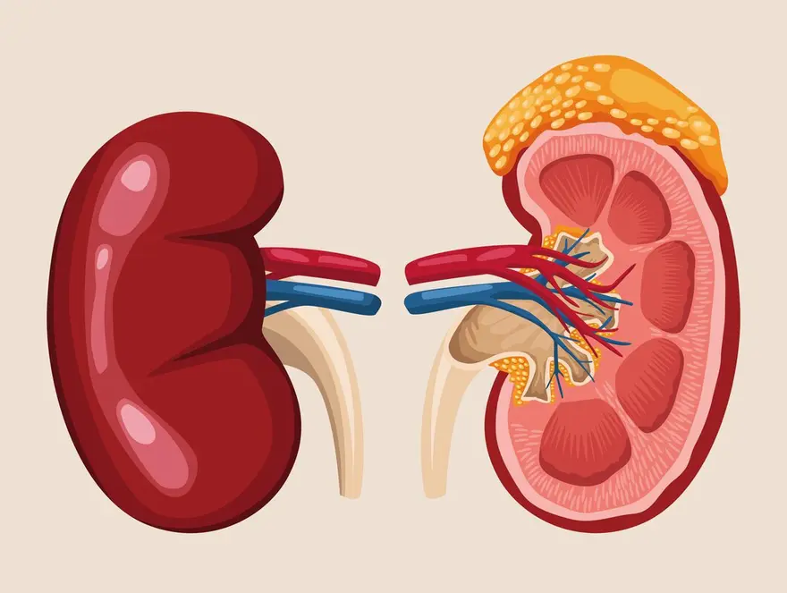
A pheochromocytoma is a rare tumour of the adrenal gland that pumps out adrenaline and related hormones. That flood of hormones drives spells of high blood pressure, headache, sweating and a pounding heart. It’s usually benign. Diagnosis starts with a blood or urine test measuring metanephrines, the breakdown products of those hormones, then a scan to locate the tumour. Getting it right before any surgery is critical.
According to Dr. Sandeep Nayak, Surgical Oncologist in India, “A pheochromocytoma is uncommon, but it’s one tumour you never want to miss. It floods the body with adrenaline, causing those dramatic spells of high blood pressure and palpitations. We diagnose it by measuring metanephrines first, then imaging to find it. The reason this matters so much is safety. Operating on an unprepared pheochromocytoma can trigger a dangerous crisis, so the diagnosis has to come first, every time.”
Having unexplained blood pressure spells and palpitations?
What Is It and What Does It Do?
Understanding this tumour means understanding the hormones it releases and the havoc they cause.
- Where it sits : It arises in the medulla, the inner part of the adrenal gland, which normally produces adrenaline in controlled amounts. This tumour makes far too much.
- The hormone surge : It pours out catecholamines, adrenaline and noradrenaline, unchecked. That surge is behind every symptom the tumour causes.
- The classic triad : Headache, sweating and palpitations occurring together, often in episodes, is the hallmark. Anxiety, tremor and pallor often join them.
- Blood pressure spells : The signature feature is high blood pressure, sometimes constant, often in sudden dramatic spikes that come and go without obvious cause.
This is one of the more demanding tumours handled within adrenal tumor treatment, precisely because of the hormones it releases.
How Is It Diagnosed?
The diagnosis follows a careful order, hormones first, then imaging, for good reason.
- Metanephrine testing : The key test measures metanephrines in blood or urine. These are the stable breakdown products of the tumour’s hormones, and they’re highly sensitive.
- Why biochemistry first : Confirming the tumour is active before imaging avoids confusion, and crucially flags the crisis risk before anyone plans surgery.
- Locating it : Once the biochemistry confirms it, a CT or MRI scan pinpoints the tumour in the adrenal gland so it can be removed safely.
- Functional imaging : In some cases a specialised scan like MIBG or a PET scan is used to find tumours sitting outside the adrenal or to check for spread.
This careful pathway fits within the wider evaluation of adrenal tumors, where a pheochromocytoma needs particularly careful handling.
Why Choose Dr. Sandeep Nayak for Pheochromocytoma Care?
Dr. Sandeep Nayak is a surgical oncologist with 24 years behind him and a fellowship in laparoscopic and robotic onco-surgery. He has treated pheochromocytomas throughout his career, and knows that with this tumour the weeks of preparation before surgery matter as much as the operation. The approach means thorough biochemical diagnosis followed by careful alpha blockade to control blood pressure, since that preparation is what turns a high risk operation into a safe one.
Pheochromocytoma is where experience genuinely saves lives. Removing one without proper hormonal preparation can trigger a cardiovascular emergency on the operating table, which is why the diagnosis and the weeks of medical preparation beforehand are non negotiable. A surgeon who understands this tumour plans for it meticulously, then removes it with minimally invasive surgery once the patient is safely prepared. That combination of caution and skill is exactly what this rare tumour demands.
Frequently Asked Questions
What is a pheochromocytoma?
A rare adrenal gland tumour that overproduces adrenaline, driving high blood pressure and palpitations.
What are its main symptoms?
Episodic headache, sweating, palpitations and high blood pressure are the classic signs.
How is pheochromocytoma diagnosed?
By measuring metanephrines in blood or urine, then locating the tumour with imaging.
Is it usually cancerous?
No. Most pheochromocytomas are benign, though a small proportion can be malignant.
References
- Pheochromocytoma diagnosis and management — National Library of Medicine
- Biochemical diagnosis of pheochromocytoma — National Library of Medicine
Disclaimer: This blog is for informational and educational purposes only and is not a substitute for professional medical advice or diagnosis.
