
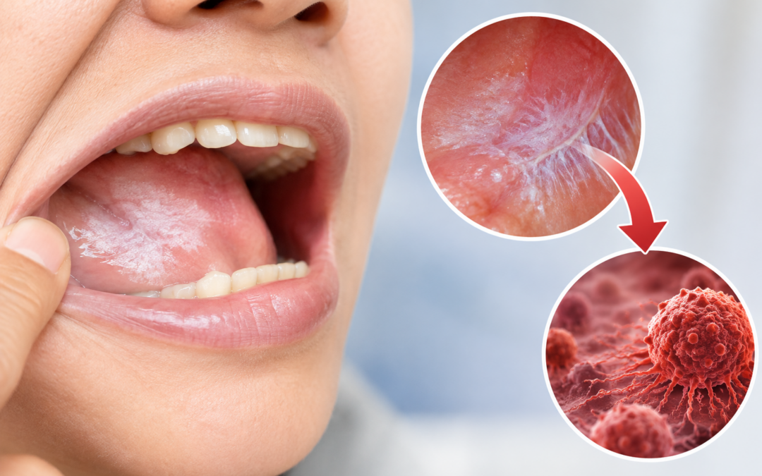
Leukoplakia and oral submucous fibrosis (OSMF) both sit on the WHO’s list of potentially malignant disorders of the mouth. That means some cases can go on to become oral squamous cell carcinoma. Most don’t. Which way a particular patient goes depends on what kind of lesion it is, where it sits, and above all whether the biopsy shows epithelial dysplasia. Here’s how to tell these conditions apart and what usually happens next.
Prof. Dr. Sandeep Nayak, Surgical Oncologist in Bengaluru at MACS Clinic, says many patients carry a white patch or a stiff mouth for months, sometimes years, simply because it doesn’t hurt. Trouble is, symptoms usually show up late, when the patch starts to burn, bleed or ulcerate. A biopsy of the affected area is the only reliable way to know whether a patch is harmless or needs treatment.
Has a white patch in your mouth been there for more than two weeks? A surgical oncologist can examine it and tell you whether a biopsy is needed.
How Likely Are Leukoplakia and Submucous Fibrosis to Turn Into Cancer?
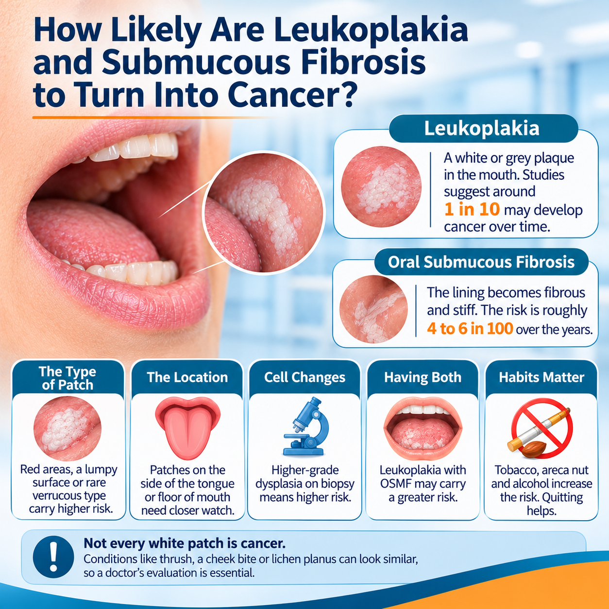
Leukoplakia. This is a white or grey plaque inside the mouth that won’t scrape off. It’s seen most often on the inner cheek (buccal mucosa), the tongue or the floor of the mouth. Pooled studies put malignant transformation at around 1 in 10 over follow-up, though the figures vary a lot from one study to the next. The blog on precancerous oral cancer stages explains how these changes are graded.
Oral submucous fibrosis. Here the lining of the cheeks and lips turns fibrous and stiff. People typically notice burning when they eat spicy food, and their mouth slowly opens less (trismus). It’s strongly linked to areca nut and gutka. Reviews estimate the risk at roughly 4 to 6 in 100 over the years.
The type of patch. A flat, evenly white plaque is lower risk. One with red or speckled areas, a lumpy surface, or the rare proliferative verrucous form is a bigger concern.
The location. Patches on the side of the tongue or the floor of the mouth get watched more closely than those on the inner cheek.
The cell changes. The biggest single factor is whether the biopsy shows epithelial dysplasia, where the surface cells mature abnormally. The higher the grade, the higher the risk.
Having both. People with leukoplakia and OSMF together have shown a higher risk than those with leukoplakia alone.
Habits matter. Tobacco in any form, areca nut and alcohol all push the risk up. Quitting brings it down.
Not every white patch is leukoplakia, of course. Thrush (candidiasis) wipes off, a cheek bite heals in a few days, and lichen planus has a lacy white pattern. It takes a doctor to sort these out.
Which White Patches in the Mouth Need a Biopsy?
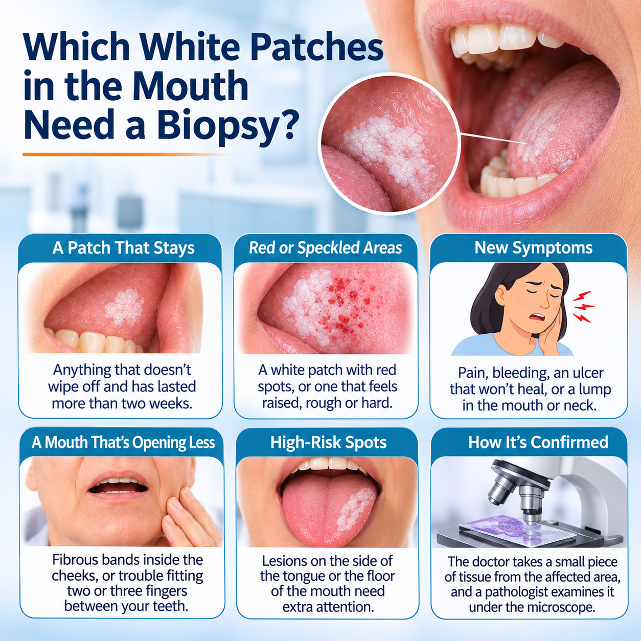
A patch that stays. Anything that doesn’t wipe off and has lasted more than two weeks.
Red or speckled areas. A white patch with red spots, or one that feels raised, rough or hard.
New symptoms. Pain, bleeding, an ulcer that won’t heal, or a lump in the mouth or neck.
A mouth that’s opening less. Fibrous bands inside the cheeks, or trouble fitting two or three fingers between your teeth.
High-risk spots. Lesions on the side of the tongue or the floor of the mouth need extra attention.
How it’s confirmed. The doctor takes a small piece of tissue from the affected area, and a pathologist examines it under the microscope. The oral cancer treatment page covers the tests involved.
What Determines the Treatment for Leukoplakia and Submucous Fibrosis?
Four things shape the plan.
The biopsy result. If there’s no dysplasia, or only mild change, the patch is usually just watched and the habit is stopped. Moderate or severe dysplasia generally means it should be removed.
The size, site and type of patch. A small, clearly bordered lesion can often be removed with surgery or a laser. A large patch, or several of them, calls for a more careful plan.
The stage of submucous fibrosis. Early disease is managed by stopping the habit, doing jaw-opening exercises and taking medicines. If mouth opening is severely restricted, surgery to release the fibrous bands may be needed.
Habits and follow-up. Giving up tobacco, areca nut and alcohol matters as much as any procedure. Follow-up visits continue for years, often every few months at first, because new lesions can appear. If cancer is found, the head and neck cancer treatment page lays out the next steps, and you’ll find more topics on the Dr. Nayak blogs page.
Key Takeaways
- Both conditions can progress to oral cancer, but most never do.
- Risk depends on the lesion’s type, its location and the dysplasia seen on biopsy.
- Reported progression is roughly 1 in 10 for leukoplakia and 4 to 6 in 100 for OSMF, with wide variation between studies.
- A white patch lasting beyond two weeks, or a mouth that opens less, should be examined.
- Quitting tobacco and areca nut and keeping up regular reviews lowers the risk.
Frequently Asked Questions
Can white patches in the mouth become cancerous?
A few can, mainly leukoplakia, but most stay benign. The risk depends on the type of lesion, where it is and whether dysplasia is present.
What is the cancer risk in oral submucous fibrosis?
About 4 to 6 in 100 patients develop oral cancer over the years, and carrying on with tobacco or areca nut raises that risk.
How do doctors confirm the cause of a white patch in the mouth?
With a clinical examination and a biopsy, which shows whether dysplastic cells are present.
Does leukoplakia regress after quitting tobacco?
Some lesions shrink or fade, but any that stay need a specialist’s review.
References
- Oral potentially malignant disorders: a consensus report from the WHO Collaborating Centre for Oral Cancer (NIH / PubMed). This report sets out the international classification of potentially malignant mouth disorders, including leukoplakia and submucous fibrosis.
- WHO: Comprehensive assessment of evidence on oral cancer prevention (World Health Organization). WHO says that avoiding tobacco, areca nut and alcohol significantly lowers the risk of oral cancer.
Disclaimer: This blog is for informational and educational purposes only and is not a substitute for professional medical advice or diagnosis.
