
Why Doesn’t Eating More Stop Cancer Weight Loss?
Cancer rewires the way the body uses food. So eating more, even of the right things, doesn’t reverse the weight loss the way it would in any other situation. Inflammation from the tumour pushes muscle to break down. Insulin resistance blocks the calories from being properly used. The result? Three full meals a day, kilos still dropping.
According to Prof. Dr. Sandeep Nayak, Surgical Oncologist in India, “Families often blame themselves when the patient keeps losing weight despite eating well. That guilt is misplaced. The problem isn’t the food, it’s that cancer has shifted the body’s metabolism. The only way to genuinely reverse the loss is treating the cancer alongside nutrition, never nutrition by itself.”
Eating more matters. It just isn’t the whole answer in cancer weight loss.
Why Does the Body Stop Using Food Properly During Cancer?
It’s not about appetite. The biology behind it runs much deeper.
- Chronic inflammation: Tumour cells leak inflammatory chemicals into the bloodstream. These chemicals confuse how muscle and fat cells use energy. Food gets eaten. The body just can’t turn it into stored weight.
- Insulin resistance: Cancer drags the body into insulin resistance. Glucose arrives, cells stay locked out. Muscle gets broken down to fill the energy gap.
- Hormone shifts: Tissue breaking down hormones outpace the tissue building ones. The balance tips toward wasting. Food intake doesn’t fix that imbalance on its own.
- Energy burns higher: Tumours steal calories at rest, competing with healthy tissue. Resting metabolism climbs. The same plate of food that used to maintain weight no longer does.
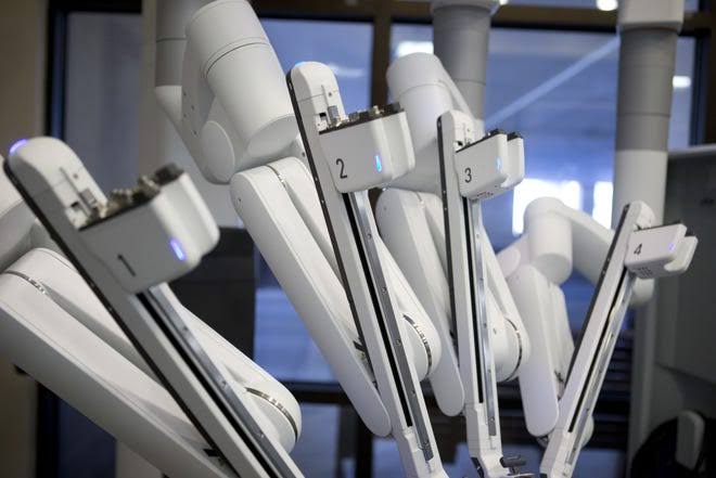
For patients whose cancer can be surgically controlled, robotic cancer surgery often slows or reverses the metabolic chaos behind the weight loss.
What Actually Helps if Eating Alone Doesn't?
Combined approaches work. Single fixes rarely do.
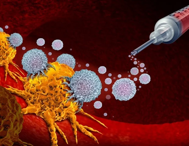
- Treat the cancer: This is the main lever. When cancer responds to chemo, targeted therapy or surgery, inflammation eases. Weight stabilises or comes back gradually.
- Medication options: Anamorelin for appetite. Megestrol for weight gain. Low dose steroids short term. Newer trial drugs like ponsegromab target the GDF15 pathway behind cachexia directly.
- Resistance exercise: Counter intuitive but proven. Light strength training holds onto muscle that calories alone can’t. Even fifteen minutes a day shows up on the scale eventually.
- Smart nutrition: Protein dense, calorie heavy. Small frequent meals, not three big ones. Oncology dietitian input where possible. Nutrition stays important, just never alone.
For patients where nutritional deficiency is also part of the picture, our blog on vitamin B12 deficiency and cancer covers another angle worth checking.
Why Choose Dr. Sandeep Nayak for Your Cancer Care?
Dr. Sandeep Nayak has spent 24 years in surgical oncology. He holds DNB qualifications in Surgical Oncology and General Surgery, plus a fellowship in Laparoscopic and Robotic Onco Surgery. He works closely with oncology dietitians, supportive care teams and medical oncologists to address cancer weight loss through combined intervention rather than nutrition alone.
Every case at MACS Clinic is reviewed by the multidisciplinary tumour board before treatment planning. Call +91 8104310753 to book your consultation.
Frequently Asked Questions
Why doesn't eating more reverse cancer weight loss?
Cancer changes metabolism so the body cannot use the extra calories.
Can a feeding tube help?
Feeding tubes help selectively, but don’t fully reverse advanced cachexia either.
What actually helps with cancer weight loss?
Treating the cancer, plus medication, exercise and nutrition together.
Does the weight come back after treatment?
Yes, partly, if the cancer responds and inflammation reduces.
Disclaimer: This blog is for informational purposes only and is not a substitute for professional medical advice.