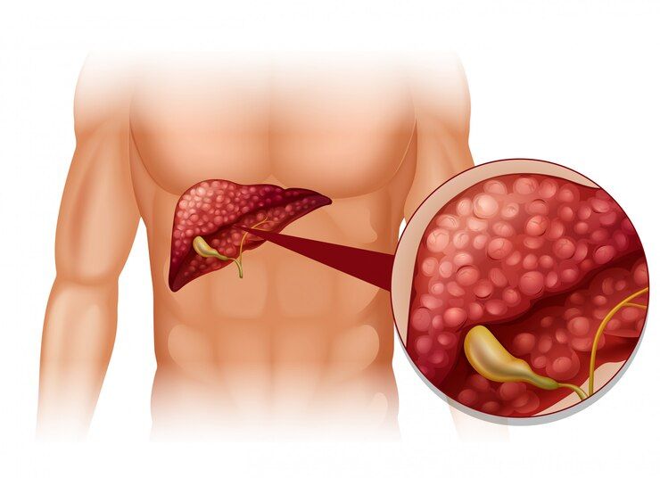
Why Do Some Cancers Spread to the Liver First?
The liver is the most common first stop for cancers that spread. It’s all about blood flow. Blood from the entire gut, pancreas and spleen flows directly into the liver through the portal vein, carrying any loose tumour cells with it. The liver also has a second blood supply from the hepatic artery. Two roads in, lots of cells getting trapped. Add the slow sinusoidal flow inside the liver and the rich growth environment, and you’ve got a perfect filter for cancer cells looking to settle.
According to Prof. Dr. Sandeep Nayak, Surgical Oncologist in India, “The liver becomes the first metastatic site for many cancers simply because of plumbing. Blood from the digestive system runs straight through it via the portal vein, so tumour cells from colon, pancreatic or stomach cancers land there first. It’s why we always image the liver carefully in any abdominal cancer staging.”
Liver mets isn’t the end, it’s where smart treatment begins.
Why Is the Liver the Most Common First Site?
A few reasons stack up together. Anatomy mostly does the rest.
- Portal vein highway: All blood draining the gut, pancreas and spleen passes through the liver first. Any cancer cell that breaks off from these organs gets carried straight in.
- Dual blood supply: The liver takes blood from two sources, the portal vein and the hepatic artery. More blood flow means more chances for circulating tumour cells to lodge.
- Slow sinusoidal flow: Blood slows down inside the liver’s tiny sinusoid channels. Cancer cells have time to stick to the lining and start growing there.
- Rich growth environment: The liver’s full of growth factors, oxygen and nutrients. Tumour cells that land here find a welcoming neighbourhood.
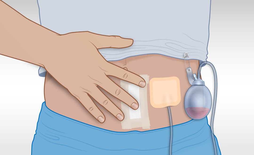
For patients diagnosed with liver mets that can be surgically removed, robotic cancer surgery brings precise resection while sparing healthy liver tissue.
Which Cancers Go to the Liver First and Why?
Pattern’s fairly predictable once you know the anatomy.
- Colorectal cancer: The classic liver-first cancer. Blood from the colon and rectum drains via the portal vein straight to the liver. About half of all colorectal patients develop liver mets.
- Pancreatic and stomach: Same portal vein route applies. Both drop liver mets early in the disease, often before other organs show involvement at all.
- Breast cancer: Goes to bone first usually, but liver is the third most common site. The liver becomes a sanctuary site once breast cancer cells settle there.
- Lung and ovarian: Lung cancer can hit the liver through the systemic circulation. Ovarian spreads to the peritoneum first, but the liver follows close behind.
For a fuller picture of how cancer spreads through different pathways, our blog on the 3 ways cancer can spread walks through direct invasion, lymphatic and bloodstream routes.
Why Choose Dr. Sandeep Nayak for Your Cancer Care?
Dr. Sandeep Nayak has spent 24 years in surgical oncology. He holds DNB qualifications in Surgical Oncology and General Surgery and trained further with a fellowship in Laparoscopic and Robotic Onco Surgery. He treats liver metastases from colorectal, pancreatic, stomach and other primary cancers with precise robotic and laparoscopic resection, alongside chemotherapy and targeted therapy planning to address both the liver lesions and the original tumour.
That combined surgical and systemic approach is what gives many liver metastasis patients realistic long term outcomes. Every case at MACS Clinic goes through tumour board review, where the treatment plan is set together. Call +91 8104310753 to book your consultation.
Frequently Asked Questions
Why do cancers spread to the liver first?
Liver receives blood directly from gut via the portal vein.
Which cancers go to liver first?
Colorectal, pancreatic, stomach, breast, lung and ovarian cancers commonly.
Are liver metastases treatable?
Yes, with surgery, chemotherapy or targeted therapy depending on case.
Can liver metastases be cured?
Some can, especially colorectal cancer with limited liver involvement.
Disclaimer: This blog is for informational purposes only and is not a substitute for professional medical advice.