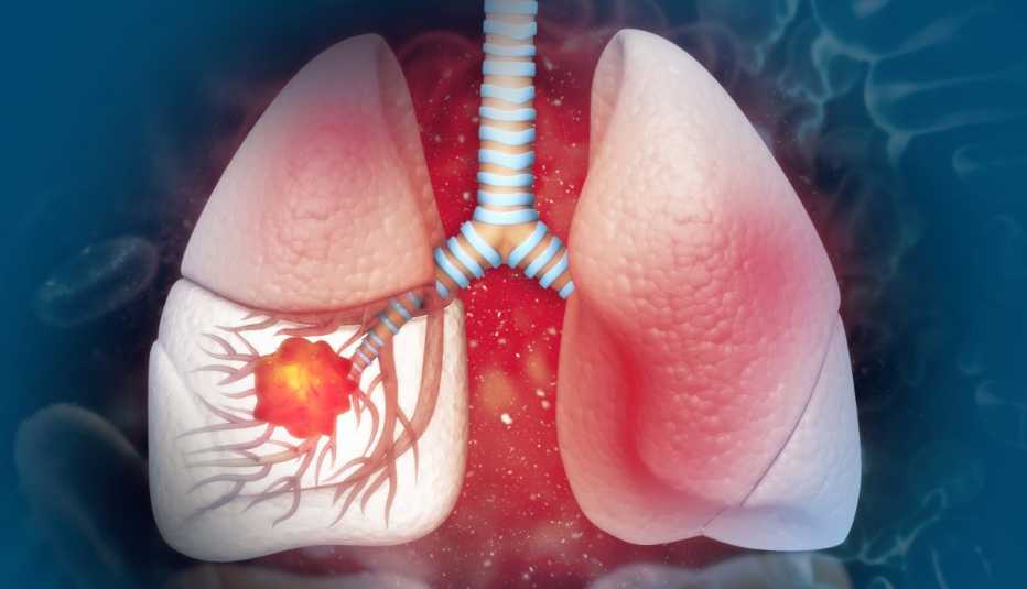
Can Kidney Cancer Be Treated Without Full Removal?

Can Kidney Cancer Be Treated Without Full Removal?

Kidney cancer under 4cm and selected tumours up to 7cm can be treated with partial nephrectomy, removing only the tumour and a clear margin of surrounding tissue while preserving the remaining functional kidney. Oncological outcomes for partial nephrectomy in T1 tumours are equivalent to radical nephrectomy removing the entire kidney. Full removal is indicated when the tumour is large, centrally located, involves the renal hilum or when partial resection cannot achieve clear margins safely. The decision is made at tumour board based on tumour size, location, the patient’s baseline kidney function and whether a minimally invasive approach is technically feasible.

According to Prof. Dr. Sandeep Nayak, Surgical Oncologist in India,
“Preserving the kidney matters beyond just the surgery. Patients with one functioning kidney face higher long-term cardiovascular and renal risk. Where partial nephrectomy is oncologically equivalent, it is the better operation.”

Have a kidney tumour and want to know whether full removal is actually necessary?

When Can Kidney Cancer Be Treated Without Full Removal?

Partial nephrectomy is now the standard of care for eligible kidney tumours and is offered at high-volume oncology centres using laparoscopic or robotic-assisted techniques.

  • Tumours Under 4cm: T1a tumours under 4cm are the strongest candidates for partial nephrectomy with oncological outcomes identical to radical nephrectomy and kidney cancer treatment at KIMS Hospital, Bangalore offers robotic-assisted partial nephrectomy for eligible patients as the preferred approach over open surgery.
  • Tumours 4 to 7cm: Selected T1b tumours between 4 and 7cm are suitable for partial nephrectomy when the tumour is exophytic, peripherally located and not involving the collecting system, with oncological equivalence to radical nephrectomy confirmed in current evidence.
  • Single Kidney Patients: Patients with a solitary kidney, bilateral kidney tumours or compromised contralateral kidney function require partial nephrectomy regardless of tumour size because radical nephrectomy in this group would leave the patient dialysis-dependent.
  • Active Surveillance Option: Very small incidentally detected tumours under 2cm in elderly patients or those with significant comorbidities are managed with active surveillance including regular imaging every three to six months rather than immediate surgery.

Where partial nephrectomy is technically achievable with clear margins, it is always preferable to radical nephrectomy from a long-term kidney function and cardiovascular risk standpoint.

When Is Full Kidney Removal Actually Necessary?

Radical nephrectomy remains the appropriate operation for specific tumour characteristics where partial resection cannot safely achieve the oncological goals of the operation.

  • Large or Central Tumours: Tumours over 7cm or those involving the renal hilum, collecting system or renal vein make partial nephrectomy technically unreliable for achieving clear margins and robotic cancer surgery or conventional radical nephrectomy is planned when the tumour anatomy makes kidney preservation oncologically unsafe.
  • Multiple Tumours: Multiple tumours distributed across the kidney make partial nephrectomy technically complex and in cases where tumour load prevents adequate remnant kidney preservation, radical nephrectomy is the safer oncological choice.
  • Locally Advanced Disease: Tumours with direct invasion into adjacent structures, perinephric fat involvement confirmed on imaging or renal vein thrombus require radical nephrectomy with extended resection rather than any kidney-preserving approach.
  • Poor Functional Remnant: When preoperative imaging and functional assessment show that the remnant kidney after partial nephrectomy would provide insufficient filtration based on the patient’s baseline function, radical nephrectomy with appropriate long-term renal management planning becomes the clinically safer option.

Tumour board review determines whether partial or radical nephrectomy is appropriate for each individual case and for more on specialist robotic kidney surgery, our blog on robotic surgery covers this in detail.

Why Choose Dr. Sandeep Nayak for Kidney Cancer Surgery ?

Dr. Sandeep Nayak brings 24 years of surgical oncology experience, DNB qualifications in Surgical Oncology and General Surgery and a fellowship in Laparoscopic and Robotic Onco-Surgery to kidney cancer surgery including robotic-assisted partial and radical nephrectomy at KIMS Hospital, Bangalore. He heads Oncology Services across Karnataka with originator credits for RABIT, MIND and L-VEIL techniques and over 25 published clinical studies. Patients with kidney tumours wanting clarity on whether full removal is necessary are seen here with every decision through tumour board review. Call +91 8104310753 to book your consultation.

Frequently Asked Questions

Can kidney cancer be cured without removing the whole kidney?

Kidney cancer under 7cm in eligible locations is treated with partial nephrectomy producing equivalent oncological outcomes to full kidney removal while preserving kidney function.

What size kidney tumour requires full removal?

Tumours over 7cm, centrally located tumours involving the renal hilum and locally advanced tumours with adjacent structure invasion typically require radical nephrectomy rather than partial resection.

Is robotic surgery available for partial nephrectomy in Bangalore?

Robotic-assisted partial nephrectomy is available at KIMS Hospital, Bangalore and is the preferred approach for eligible kidney tumours over open or conventional laparoscopic surgery.

What happens to kidney function after partial nephrectomy?

Partial nephrectomy preserves the functional remnant kidney, maintaining better long-term renal function and reducing cardiovascular risk compared to radical nephrectomy removing the entire kidney.

Reference Links-

  1. National Cancer Institute — Kidney Cancer Treatment
  2. World Health Organization — Renal Cell Carcinoma
  • Disclaimer: The information shared in this content is for educational purposes and not for promotional use.
First Symptoms of Liver Cancer in Indians

First Symptoms of Liver Cancer in Indians

In Indians, the first symptoms of liver cancer are upper right abdominal discomfort, unexplained weight loss, persistent fatigue and loss of appetite. These appear in a liver already damaged by Hepatitis B, Hepatitis C or alcohol-related cirrhosis, which is why they get blamed on the underlying disease rather than a new tumour developing within it. Jaundice and abdominal swelling appear later and by that point the disease is usually advanced.

According to Prof. Dr. Sandeep Nayak, Surgical Oncologist in India,
“Most liver cancer patients arrive with symptoms that started months earlier. The challenge is those early symptoms look identical to the cirrhosis they already have. That’s why surveillance in high-risk patients is not optional.”

Have known liver disease and want a specialist liver cancer assessment?

What Are the First Symptoms of Liver Cancer in Indians?

Early liver cancer symptoms are non-specific and consistently get attributed to existing liver disease rather than a new malignancy.

  • Right Abdominal Discomfort: A dull ache or heaviness in the upper right abdomen that is new or has changed character from the patient’s existing liver disease symptoms is the most common early presenting complaint in hepatocellular carcinoma and liver cancer treatment assessment at KIMS Hospital, Bangalore begins with AFP testing and dedicated liver imaging for any such new symptom in a high-risk patient.
  • Unexplained Weight Loss: Weight loss without dietary change in a patient with known chronic liver disease is a specific red flag that requires investigation rather than being attributed to poor appetite from cirrhosis alone.
  • Loss of Appetite: Persistent anorexia that worsens progressively over weeks rather than fluctuating with liver disease activity is a symptom that distinguishes developing hepatocellular carcinoma from stable compensated cirrhosis in most clinical presentations.
  • Persistent Fatigue: Fatigue that is new, worsening or disproportionate to the patient’s known liver disease severity warrants AFP and ultrasound review rather than reassurance, particularly in patients with known Hepatitis B or C infection.

These four symptoms in any patient with chronic liver disease require immediate AFP testing and liver ultrasound, not watchful waiting.

What Later Symptoms Indicate Liver Cancer Has Progressed?

When these symptoms appear, the disease has typically moved beyond the stage where curative resection is straightforward.

  • Jaundice: Yellow discolouration of the skin and eyes in a liver cancer patient indicates biliary obstruction from tumour growth or advancing liver failure and robotic cancer surgery or conventional liver resection is significantly more complex when jaundice is present at the time of surgical assessment.
  • Abdominal Swelling: Ascites, fluid accumulation in the abdomen, reflects deteriorating liver function and portal hypertension from advancing tumour burden and its appearance signals that the disease has moved well beyond early-stage resectable hepatocellular carcinoma.
  • Palpable Abdominal Mass: A lump felt in the upper right abdomen indicates a tumour large enough to be palpable through the abdominal wall, which in most cases corresponds to a tumour size that significantly complicates or precludes curative surgical resection.
  • Shoulder Tip Pain: Right shoulder tip pain referred from an enlarging right lobe hepatic tumour irritating the diaphragm is a specific symptom of advanced hepatocellular carcinoma that patients frequently attribute to musculoskeletal causes for weeks before the correct investigation is ordered.

Surveillance with six-monthly AFP and liver ultrasound in all high-risk patients is what detects liver cancer before these later symptoms appear and for more on how cancer diagnosis is confirmed, our blog on cancer diagnosis covers this in detail.

Why Choose Dr. Sandeep Nayak for Liver Cancer Surgery ?

Dr. Sandeep Nayak brings 24 years of surgical oncology experience, DNB qualifications in Surgical Oncology and General Surgery and a fellowship in Laparoscopic and Robotic Onco-Surgery to liver cancer surgery including laparoscopic and robotic-assisted hepatic resection at KIMS Hospital, Bangalore. He heads Oncology Services across Karnataka with originator credits for RABIT, MIND and L-VEIL techniques and over 25 published clinical studies. Patients with known liver disease, abnormal AFP or suspected hepatocellular carcinoma are seen here with every case reviewed through tumour board. Call +91 8104310753 to book your consultation.

Frequently Asked Questions

What are the first signs of liver cancer in Indians?

Upper right abdominal discomfort, unexplained weight loss, loss of appetite and persistent fatigue are the most common first symptoms, all of which get attributed to existing liver disease.

Who is at highest risk of liver cancer in India?

Patients with chronic Hepatitis B, Hepatitis C or alcohol-related cirrhosis carry the highest risk and require six-monthly AFP testing and liver ultrasound surveillance.

Does liver cancer cause jaundice early?

Jaundice appears in advanced liver cancer rather than early disease and indicates significant tumour burden or biliary obstruction, both of which complicate surgical management.

Can liver cancer be cured if caught early?

Surgically resectable hepatocellular carcinoma detected through surveillance at an early stage carries a five-year survival rate significantly higher than cancer diagnosed from symptoms alone.

Reference Links-

  1. National Cancer Institute — Liver Cancer Symptoms and Diagnosis
  2. World Health Organization — Hepatocellular Carcinoma
  • Disclaimer: The information shared in this content is for educational purposes and not for promotional use.
Early Signs of Stomach Cancer Most People Miss

Early Signs of Stomach Cancer Most People Miss

Stomach cancer is one of the most commonly missed cancers in India because its early symptoms are identical to ordinary indigestion. Persistent upper abdominal discomfort, early satiety, mild nausea and unexplained weight loss are the four signs patients most frequently attribute to acidity, stress or dietary habits for months before seeking investigation. By the time the diagnosis is confirmed, most cases in India are at Stage 3 or Stage 4. The signs listed below are individually non-specific but in combination, especially in patients over 45 with a family history or H. pylori infection, they warrant urgent endoscopic evaluation rather than empirical antacid therapy.

According to Prof. Dr. Sandeep Nayak, Surgical Oncologist in India,
“The patients who reach us early are almost always those whose general physician didn’t treat persistent upper abdominal symptoms with antacids alone. They investigated first. That decision is what changes the outcome.”

Experiencing persistent upper abdominal symptoms and want a specialist assessment?

What Are the Early Signs of Stomach Cancer That Get Dismissed?

The symptoms of early stomach cancer are almost never dramatic. They look exactly like common gut complaints and that is precisely why they get missed for so long.

  • Persistent Indigestion: Indigestion or heartburn that doesn’t improve with antacids after two to three weeks or returns consistently after stopping medication is one of the most overlooked early indicators and stomach and esophageal cancer assessment at KIMS Hospital, Bangalore begins with urgent endoscopy for anyone over 45 presenting with new-onset persistent dyspepsia.
  • Early Satiety: Feeling full after eating only a small amount is a symptom patients consistently normalise as poor appetite or stress, but early satiety that appears without a dietary change and persists for more than two to three weeks is a red flag for gastric tumour causing reduced stomach capacity.
  • Unexplained Weight Loss: Losing weight without intentional dietary change or increased activity is a systemic cancer symptom that applies across multiple cancer types and unexplained weight loss of more than 5 percent of body weight over six months warrants investigation regardless of how benign the patient’s other symptoms appear.
  • Upper Abdominal Discomfort: A vague ache or pressure in the upper abdomen that is not clearly related to meals, doesn’t respond to antacids and persists across several weeks is consistently described by patients with early gastric cancer as something they dismissed for months before it worsened.

These four symptoms together in a patient over 45 with H. pylori history, a family history of stomach cancer or a diet high in smoked and salted foods constitute a clinical indication for immediate endoscopy.

What Other Signs Should Raise Concern for Stomach Cancer?

Several additional signs appear slightly later in the early disease process and are still actionable if investigated promptly.

  • Nausea Without Cause: Persistent low-grade nausea without a clear dietary or medication trigger, particularly when it appears alongside early satiety or upper abdominal discomfort, is a combination that warrants endoscopic investigation rather than empirical antiemetic treatment.
  • Blood in Stool or Vomit: Vomiting blood or passing dark tarry stools indicates bleeding from the upper gastrointestinal tract and robotic cancer surgery or conventional gastric resection for surgically identified stomach cancer produces significantly better outcomes when the disease is caught before this symptom appears.
  • Difficulty Swallowing: Dysphagia involving solid foods progressing to softer foods is a specific symptom of tumours at the gastro-oesophageal junction, the area where the stomach meets the oesophagus, and this symptom should never be attributed to acid reflux without endoscopic confirmation.
  • Anaemia Without Explanation: Iron deficiency anaemia without a clear source of blood loss in a patient over 45 requires upper and lower gastrointestinal investigation because chronic slow bleeding from an early gastric tumour is a common presentation that gets managed as dietary anaemia for months before the correct diagnosis is made.

Early stomach cancer is treatable and surgically curable. The problem is never the surgery. It is how long the diagnosis takes and for more on how early cancer signs are investigated, our blog on early signs of cancer covers the investigation approach in detail.

Why Choose Dr. Sandeep Nayak for Stomach Cancer Surgery ?

Dr. Sandeep Nayak brings 24 years of surgical oncology experience, DNB qualifications in Surgical Oncology and General Surgery and a fellowship in Laparoscopic and Robotic Onco-Surgery to stomach cancer surgery including laparoscopic and robotic-assisted gastrectomy at KIMS Hospital, Bangalore. He heads Oncology Services across Karnataka with originator credits for RABIT, MIND and L-VEIL techniques and over 25 published clinical studies. Patients with persistent upper GI symptoms or a confirmed stomach cancer diagnosis are seen here with every case reviewed through tumour board. Call +91 8104310753 to book your consultation.

Frequently Asked Questions

What are the earliest signs of stomach cancer?

Persistent indigestion, early satiety, unexplained weight loss and upper abdominal discomfort are the most common early signs that patients dismiss as ordinary gut problems for months.

At what age should stomach cancer symptoms be investigated urgently?

New-onset persistent upper abdominal symptoms in anyone over 45, particularly with H. pylori history or family history of gastric cancer, warrant urgent endoscopy rather than empirical antacid treatment.

Can stomach cancer cause anaemia?

Chronic slow bleeding from an early gastric tumour frequently presents as unexplained iron deficiency anaemia and this is one of the most commonly missed indirect indicators of early stomach cancer.

Is stomach cancer curable if caught early?

Early-stage stomach cancer is surgically curable and resection at Stage 1 or Stage 2 produces five-year survival rates significantly higher than surgery performed at Stage 3 or Stage 4.

Reference Links-

  1. National Cancer Institute — Stomach Cancer Symptoms and Diagnosis
  2. World Health Organization — Gastric Cancer
  • Disclaimer: The information shared in this content is for educational purposes and not for promotional use.
Lumpectomy vs Mastectomy Cost in Bangalore 2025

Lumpectomy vs Mastectomy Cost in Bangalore 2025

Lumpectomy in Bangalore costs INR 75,000 to 2,00,000 at private specialist centres. Mastectomy runs INR 1,00,000 to 3,50,000 with modified radical mastectomy at the higher end. These figures cover surgery, anaesthesia, theatre and a two to four day hospital stay. Lumpectomy is the smaller operation but it always requires radiation afterward, which adds INR 90,000 to 3,50,000 to the total. Mastectomy costs more upfront but avoids mandatory radiation in many cases. The right choice between the two is never purely financial. It’s driven by tumour size, margin achievability and what the tumour board recommends for that specific case.

According to Prof. Dr. Sandeep Nayak, Surgical Oncologist in India,
“Lumpectomy and mastectomy produce equivalent survival in appropriately selected patients. The cost difference between them is real but it should never be the deciding factor. The oncological decision comes first.”

Want a clear cost estimate for your specific breast cancer surgical plan in Bangalore?

What Does Lumpectomy Cost in Bangalore in 2025?

Lumpectomy is the less expensive operative procedure but the full treatment cost including mandatory radiation is higher than the surgical fee alone.

  • Operative Cost: Lumpectomy at private specialist centres in Bangalore costs INR 75,000 to 2,00,000 covering the operation, anaesthesia, theatre charges and a two day hospital stay and breast cancer treatment at high-volume oncology centres like KIMS sits toward the upper end of that range.
  • Radiation Is Mandatory: Every lumpectomy requires radiation to the remaining breast tissue and a complete course in Bangalore costs INR 90,000 to 3,50,000, making the total lumpectomy plus radiation cost INR 1,65,000 to 5,50,000 at private specialist centres.
  • Sentinel Node Biopsy Added: Axillary staging through sentinel node biopsy runs in the same session at an additional INR 40,000 to 80,000 and the result directly determines whether adjuvant chemotherapy is added to the treatment plan after surgery.
  • Oncoplastic Lumpectomy Premium: Oncoplastic techniques that reshape the breast at the time of tumour removal add a surgical premium of INR 30,000 to 80,000 over standard lumpectomy at centres with dedicated oncoplastic breast surgery capability in Bangalore.

Lumpectomy’s lower operative cost is partially offset by the mandatory radiation course that follows every breast-conserving procedure regardless of tumour biology or stage.

Lumpectomy vs Mastectomy Cost Comparison in Bangalore 2025

Lumpectomy

Mastectomy

Operative Cost

INR 75,000 to 2,00,000

INR 1,00,000 to 3,50,000

Hospital Stay

1 to 2 days

3 to 5 days

Radiation Required

Always

Selective at Stage 1 and 2

Reconstruction

Not usually needed

Optional, adds INR 1 to 5 lakhs

Total Surgery Cost

INR 1.65 to 5.5 lakhs with radiation

INR 1 to 8.5 lakhs with reconstruction

Recovery Time

2 to 3 weeks

4 to 6 weeks

  • Mastectomy Without Reconstruction: Simple or modified radical mastectomy at private specialist centres in Bangalore costs INR 1,00,000 to 3,50,000 covering surgery, anaesthesia, theatre and a three to five day hospital stay and robotic cancer surgery or conventional mastectomy packages at KIMS Hospital are quoted as all-inclusive figures before any procedure is booked.
  • Reconstruction Adds Significantly: Immediate implant-based reconstruction adds INR 1,00,000 to 2,50,000 while flap reconstruction adds INR 2,00,000 to 5,00,000 on top of the mastectomy fee, making total mastectomy plus reconstruction cost INR 2 to 8.5 lakhs depending on technique.
  • Post-Mastectomy Radiation at Stage 3: Mastectomy avoids radiation in most Stage 1 and 2 cases but Stage 3 disease requires post-mastectomy chest wall radiation regardless, adding the same INR 90,000 to 3,50,000 radiation cost to both procedures at locally advanced stages.
  • Long-Term Cost Is Similar: When radiation after lumpectomy and reconstruction after mastectomy are both factored in, the total treatment cost difference between the two procedures at private specialist centres in Bangalore is smaller than the operative fee gap suggests.

The right operation is determined by oncological criteria not cost and for more on what qualifies a patient for breast conservation, our blog on breast conserving surgery covers this in detail.

Why Choose Dr. Sandeep Nayak for Breast Cancer Treatment?

Dr. Sandeep Nayak brings 24 years of surgical oncology experience, DNB qualifications in Surgical Oncology and General Surgery and a fellowship in Laparoscopic and Robotic Onco-Surgery to lumpectomy, mastectomy and oncoplastic breast surgery at KIMS Hospital, Bangalore. He heads Oncology Services across Karnataka with originator credits for RABIT and over 25 published clinical studies. Patients wanting a transparent cost breakdown alongside a clear surgical recommendation are seen here with every decision going through tumour board review. Call +91 8104310753 to book your consultation.

Frequently Asked Questions

How much does lumpectomy cost in Bangalore in 2025?

Lumpectomy at private specialist centres in Bangalore costs INR 75,000 to 2,00,000 covering surgery, anaesthesia, theatre charges and a two day hospital stay.

How much does mastectomy cost in Bangalore in 2025?

Mastectomy costs INR 1,00,000 to 3,50,000 at private specialist centres with modified radical mastectomy at the higher end depending on operative complexity and axillary surgery extent.

Is lumpectomy cheaper than mastectomy overall?

Lumpectomy has a lower operative fee but mandatory radiation adds INR 90,000 to 3,50,000 making the total treatment cost comparable to mastectomy without reconstruction.

Does mastectomy always require reconstruction in Bangalore?

Reconstruction after mastectomy is optional and patient-driven. Implant reconstruction adds INR 1 to 2.5 lakhs and flap reconstruction adds INR 2 to 5 lakhs on top of the mastectomy cost.

Reference Links-

  1. National Cancer Institute — Breast Cancer Surgery Options
  2. Medijourney — Breast Cancer Treatment Cost Bangalore
  • Disclaimer: The information shared in this content is for educational purposes and not for promotional use.
How Is Pancreatic Cancer Diagnosed Early

How Is Pancreatic Cancer Diagnosed Early

Pancreatic cancer is one of the hardest cancers to detect early because the pancreas sits deep in the abdomen and produces no reliable symptoms until the tumour is large or has spread. Most cases are diagnosed at Stage 3 or Stage 4. Early diagnosis happens in two specific situations: incidental detection on imaging ordered for another reason or systematic surveillance in patients with confirmed high-risk factors including hereditary pancreatitis, BRCA2 mutation, familial pancreatic cancer syndrome or longstanding Type 2 diabetes with unexplained weight loss.

According to Prof. Dr. Sandeep Nayak, Surgical Oncologist in India,
“Pancreatic cancer found early is surgically curable. The problem is the biology of the disease, not the surgery. Most patients don’t have symptoms until the window for curative resection has already closed.”

Want a specialist assessment for pancreatic cancer risk or an incidental pancreatic finding?

What Tests Are Used to Diagnose Pancreatic Cancer Early?

Early diagnosis relies on imaging, tumour markers and biopsy in combination rather than any single test used alone.

  • CT Pancreas Protocol: A dedicated pancreatic phase CT scan is the primary imaging tool for identifying pancreatic tumours, assessing vascular involvement and staging resectability and pancreatic and bile duct cancer assessment at KIMS Hospital, Bangalore uses this as the first-line investigation for any suspected pancreatic lesion.
  • Endoscopic Ultrasound: EUS provides higher resolution imaging of the pancreatic head, body and tail than CT alone and allows fine needle aspiration biopsy of lesions too small to sample by percutaneous route, making it essential for diagnosing small tumours and cystic lesions.
  • CA 19-9 Tumour Marker: CA 19-9 is elevated in most pancreatic cancers but is not specific enough to use as a standalone screening test because it is also elevated in bile duct obstruction, pancreatitis and other benign conditions, making it most useful alongside imaging rather than alone.
  • MRCP and PET Scan: MRCP defines ductal anatomy and identifies strictures or cystic lesions that may indicate early malignant change while PET-CT identifies metabolically active disease and confirms or excludes distant spread before any surgical decision is made.

A confirmed early-stage pancreatic tumour on imaging requires biopsy for histological diagnosis before any treatment plan is confirmed.

Who Is at High Risk and Needs Active Surveillance for Pancreatic Cancer?

Systematic surveillance in high-risk individuals is currently the most clinically reliable pathway to early pancreatic cancer diagnosis.

  • Hereditary Pancreatitis: Patients with confirmed hereditary pancreatitis have a 40 to 75 times higher lifetime risk of pancreatic cancer and annual EUS or MRI surveillance from age 40 is the clinical standard for this group, not a precautionary option.
  • BRCA2 and PALB2 Mutations: Confirmed BRCA2 or PALB2 mutation carriers with a first-degree relative with pancreatic cancer are recommended annual EUS and MRI surveillance from age 50 or ten years before the youngest affected relative’s diagnosis age and robotic cancer surgery for resectable pancreatic lesions identified through surveillance produces significantly better outcomes than surgery for symptomatic disease.
  • New Onset Diabetes Over 50: New-onset Type 2 diabetes in someone over 50 with unexplained weight loss and no family history of diabetes carries a clinically meaningful association with pancreatic cancer and warrants a dedicated pancreatic CT before attributing it to metabolic disease alone.
  • Intraductal Papillary Mucinous Neoplasms: IPMNs detected incidentally on abdominal imaging are premalignant lesions that require structured surveillance with EUS or MRI every six to twelve months depending on size and morphology to detect malignant transformation before it becomes invasive.

Active surveillance in high-risk groups is where early pancreatic cancer diagnosis is actually achieved and for more on how cancer biopsy confirms diagnosis, our blog on cancer diagnosis covers this in detail.

Why Choose Dr. Sandeep Nayak for Breast Cancer Treatment?

Dr. Sandeep Nayak brings 24 years of surgical oncology experience, DNB qualifications in Surgical Oncology and General Surgery and a fellowship in Laparoscopic and Robotic Onco-Surgery to pancreatic cancer surgery including Whipple procedure, distal pancreatectomy and robotic-assisted resection at KIMS Hospital, Bangalore. He heads Oncology Services across Karnataka with originator credits for RABIT, MIND and L-VEIL techniques and over 25 published clinical studies. Patients with pancreatic lesions, incidental findings or high-risk surveillance needs are seen here with every case reviewed through tumour board. Call +91 8104310753 to book your consultation.

Frequently Asked Questions

Can pancreatic cancer be detected before symptoms appear?

Pancreatic cancer can be detected before symptoms appear through systematic EUS and MRI surveillance in confirmed high-risk individuals including BRCA2 carriers and hereditary pancreatitis patients.

What is the best test to detect pancreatic cancer early?

Endoscopic ultrasound combined with dedicated pancreatic phase CT is the most reliable combination for detecting early pancreatic cancer and assessing surgical resectability.

Is CA 19-9 a reliable early detection test for pancreatic cancer?

CA 19-9 is not reliable as a standalone screening test because it is elevated in benign conditions but is clinically useful alongside imaging for staging and treatment monitoring.

Who should have regular surveillance for pancreatic cancer?

Patients with hereditary pancreatitis, confirmed BRCA2 or PALB2 mutations with family history and those with IPMN lesions require structured annual EUS or MRI surveillance.

Reference Links-

  1. National Cancer Institute — Pancreatic Cancer Screening
  2. World Health Organization — Cancer Early Detection
  • Disclaimer: The information shared in this content is for educational purposes and not for promotional use.