Dr. Sandeep Nayak treats the full range of solid tumour cancers at MACS Clinic in Bangalore and the list is longer than most patients expect when they first call because he’s spent 24 years building a practice that covers colorectal, thyroid, head and neck, gastric, liver, kidney, prostate, ovarian, adrenal and lung cancers rather than narrowing down to two or three types and referring everything else out the way most surgical oncology practices in India actually work.
According to Prof. Dr. Sandeep Nayak,Surgical Oncologist in India, “The cancers we treat at MACS Clinic cover the full surgical oncology spectrum and the approach for each one gets decided by what the individual case needs rather than by what we find most convenient to offer.”
Which Cancers Does Dr. Sandeep Nayak Treat at MACS Clinic?
These are the main cancer types Dr. Nayak operates on at MACS Clinic:
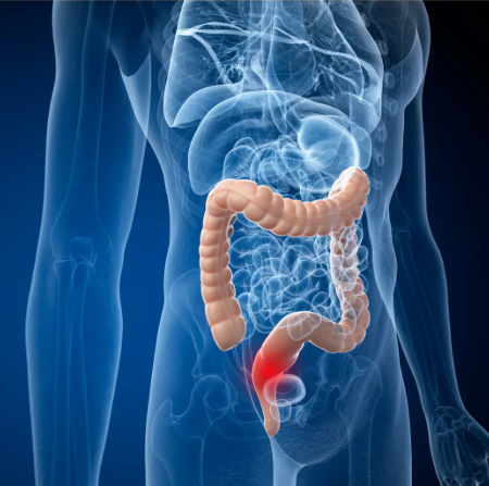
Colorectal cancer: Colon and rectal cancer with D3 resection, robotic low anterior resection, inter-sphincteric resection for stoma avoidance and HIPEC for peritoneal spread, all done laparoscopically or robotically because that’s what the evidence says patients deserve not because it sounds better on a website.
Thyroid cancer: Total thyroidectomy with neck dissection and RABIT for patients who don’t want to wake up with a scar across their neck, which Dr. Nayak built himself and performs at a volume nobody else in India is close to matching.
Head and neck cancers: Oral cancer, tongue cancer, oropharyngeal tumours, laryngeal cancer and salivary gland disease with TORS for base of tongue cases and neck dissection built into every appropriate operation from the start rather than planned separately after someone’s already been through surgery once.
Abdominal and pelvic cancers: Gastric, liver, kidney, adrenal, prostate, ovarian, uterine and pancreatic cancers all treated at MACS Clinic with a surgical approach that comes from looking at your specific scans, your specific pathology and your specific anatomy rather than from a protocol someone wrote for a different patient.
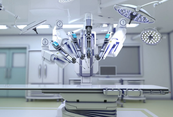
Patients who come to MACS Clinic don’t spend weeks being passed between specialists before anyone actually talks to them about surgery, they sit in front of Dr. Nayak who has operated on all of these cancer types himself at real volume and they get an assessment from someone who knows the full picture.Robotic cancer surgery at MACS Clinic covers the full minimally invasive range across all these cancers rather than limiting it to the handful of procedures most centres have committed to doing robotically.
What Makes Cancer Treatment at MACS Clinic Different?
These are the things patients who’ve been elsewhere consistently say they noticed at MACS Clinic that they hadn’t found before:
Dr. Nayak is the one who sees you: Not a junior doctor who takes notes and summarises for someone who’ll read them later, Dr. Nayak looks at your imaging himself, reads your pathology himself and has the real conversation with you himself and that sounds obvious but it genuinely isn’t what most patients experience before they get to MACS Clinic.
Minimally invasive is where every conversation starts: Nobody at MACS Clinic asks whether laparoscopic or robotic surgery is worth considering for your case, they ask whether anything about your case makes it impossible and most of the time the answer is nothing does.
The techniques are here because he built them: RABIT, MIND and RIA-MIND exist at MACS Clinic because Dr. Nayak created them here from what he learned operating at real volume and patients who need these approaches get them from the surgeon who developed them rather than from someone who read about them afterward.
Second opinions that are actually useful: Patients who’ve been told what their treatment will be and feel unsettled about it come to MACS Clinic and get told honestly whether what they were offered makes sense for their specific case or whether something else fits better and nobody in that conversation feels the need to defend what another centre decided.
Whether your cancer is on Dr. Nayak’s list and what approach your case actually calls for is worth a conversation before you agree to treatment anywhere because the difference between a plan that came from your scans and one that came from a protocol is something you feel even when you can’t articulate exactly why.Laparoscopic cancer surgery at MACS Clinic covers the full minimally invasive spectrum where every surgical decision comes from what your specific cancer needs.
Why Choose Dr. Sandeep Nayak for Cancer Treatment?
Colorectal. Thyroid. Head and neck. Gastric. Liver. Kidney. Prostate. Ovarian. Adrenal. Pancreatic. Lung. All of them, at real volume, for 24 years. Over a thousand robotic cancer surgeries. RABIT, MIND and RIA-MIND built here not borrowed. Chairman of Oncology Services across Karnataka. Kidwai Memorial Institute of Oncology alumnus. MACS Clinic Jayanagar Bangalore, Monday to Saturday 3pm to 6:30pm, plus 91 9482202240. You call, you get seen, you sit across from Dr. Nayak himself and you find out what your cancer actually needs rather than what the centre near you is most comfortable offering.
Frequently Asked Questions
What cancers does Dr. Sandeep Nayak treat at MACS Clinic? Colorectal, thyroid, head and neck, gastric, liver, kidney, prostate, ovarian, uterine, adrenal, pancreatic and lung cancers among other solid tumours at MACS Clinic Jayanagar Bangalore.
Does Dr. Sandeep Nayak treat all stages of cancer at MACS Clinic? Yes, from early stage through locally advanced and stage four including HIPEC for peritoneal spread, MACS Clinic covers the full staging spectrum.
Can I get a second opinion from Dr. Sandeep Nayak at MACS Clinic? Yes, patients with an existing diagnosis or treatment plan who want an honest case-specific assessment rather than a generic second opinion are seen at MACS Clinic.
How do I book a consultation at MACS Clinic with Dr. Sandeep Nayak? Call plus 91 9482202240, MACS Clinic Jayanagar Bangalore, Monday to Saturday 3pm to 6:30pm.
What makes Dr. Sandeep Nayak different from other oncologists in India isn’t a longer list of credentials or a fancier hospital affiliation, it’s that he spent fifteen years operating at the frontier of robotic and laparoscopic cancer surgery when most Indian centres were still deciding whether it was worth pursuing, built techniques that didn’t exist before he made them and consistently tells patients what their specific case actually needs rather than what fits most conveniently into the surgical programme the centre already runs.
According to Prof. Dr. Sandeep Nayak,Surgical Oncologist in India, “Every cancer patient deserves a surgical plan built around their specific tumour, their anatomy and their life rather than a protocol that was designed for the average case and applied to everyone.”
What Sets Dr. Sandeep Nayak Apart Technically?
These are the things that separate Dr. Nayak’s surgical practice from most oncologists operating in India today:
He built the techniques: RABIT, MIND and RIA-MIND didn’t come from a fellowship overseas or a paper someone handed him at a conference, he developed them himself from operating at real volume and paying close enough attention to outcomes that he could see where standard technique was leaving patients with less than they deserved.
Fifteen years ahead: Dr. Nayak got into robotic and laparoscopic cancer surgery more than fifteen years ago when the honest answer is most Indian oncology centres hadn’t made up their minds about it yet and that head start built a depth of experience that centres adopting it more recently are genuinely still catching up to.
Scarless thyroid surgery: RABIT removes the thyroid through the armpit and below the collarbone with no cut on the neck and for a patient who would otherwise carry a visible scar across their throat for decades that’s not a surgical preference it’s a completely different life experience after treatment.
Sphincter preservation at volume: Working deep in the narrow pelvis with the precision that inter-sphincteric rectal resection demands and doing it repeatedly enough to have built genuine mastery is what lets Dr. Nayak offer stoma avoidance in cases where a less experienced team would default to a permanent bag as the path of least resistance.
The technical depth Dr. Nayak has across specific cancer types at real volume is what separates his practice from an oncologist who operates broadly without the same depth in any of them and patients who’ve had consultations elsewhere consistently notice that difference in how the conversation about their case actually goes.Robotic cancer surgery at MACS Clinic is what fifteen years of building that depth actually looks like in practice.
What Sets Dr. Sandeep Nayak Apart as a Clinician?
These are the things patients consistently say set their experience with Dr. Nayak apart from other oncologists they’d seen before coming to MACS Clinic:
Honest over reassuring: Dr. Nayak tells patients what the surgical options genuinely are for their specific tumour rather than what sounds most encouraging and patients who’ve had consultations elsewhere where the conversation felt vague or optimistic in a way that didn’t quite land notice the difference at MACS Clinic immediately.
Your case not the last one: Every patient gets a plan built from their actual imaging, their pathology, their anatomy and their circumstances rather than a standard protocol adjusted slightly because the previous ten patients with something similar all got the same thing.
He tells you what doesn’t need operating on: Some of the most important things Dr. Nayak does in consultation are the recommendations not to operate, to monitor instead, to reassess in three months, and patients who came in expecting to be pushed toward surgery often leave with a non-surgical recommendation nobody else had offered them.
The conversation makes sense: Patients walking out of a consultation with Dr. Nayak understand what’s happening to them, why the plan is what it is and what the realistic outcomes look like and that clarity about their own situation is something a lot of them say they hadn’t had anywhere else.
What patients remember isn’t just the surgical outcome, it’s that they understood their own case before they agreed to anything and felt like the plan was built for them specifically rather than fitted around what the centre was most comfortable offering.Laparoscopic cancer surgery at MACS Clinic covers the full minimally invasive spectrum where every surgical decision starts from what your cancer actually needs.
Why Choose Dr. Sandeep Nayak for Cancer Treatment?
Over a thousand robotic cancer surgeries. Twenty four years in surgical oncology. RABIT, MIND and RIA-MIND built from operating at real volume not borrowed from anyone. Chairman of Oncology Services across Karnataka. Kidwai Memorial Institute of Oncology alumnus. Seeing patients at MACS Clinic in Jayanagar Bangalore Monday to Saturday 3pm to 6:30pm. Contact plus 91 9482202240. Dr. Nayak is the kind of surgeon who’s been doing this long enough and at enough volume that he can tell you honestly what your case needs and what it doesn’t and patients who’ve spent time in consultations that didn’t give them that clarity understand immediately why that matters.
Frequently Asked Questions
What makes Dr. Sandeep Nayak different from other oncologists? He developed original techniques including RABIT, MIND and RIA-MIND, adopted robotic cancer surgery over 15 years ago and builds every treatment plan around the specific patient in front of him rather than a standard protocol.
What techniques has Dr. Sandeep Nayak developed that other oncologists haven’t? RABIT for scarless thyroid surgery with no neck incision, MIND for robotic pelvic cancer dissection and RIA-MIND for complex low rectal cases, all built from his own operating experience at real volume.
Does Dr. Sandeep Nayak offer second opinions for cancer surgery in Bangalore? Yes, patients who want a second opinion on their cancer diagnosis or proposed treatment plan are seen at MACS Clinic and given an honest assessment of what their specific case actually requires.
Where can I consult Dr. Sandeep Nayak in Bangalore? MACS Clinic Jayanagar Bangalore, Monday to Saturday 3pm to 6:30pm, contact plus 91 9482202240.
Dr. Sandeep Nayak has performed over a thousand robotic cancer surgeries and the reason that number matters isn’t because it looks good on a website, it’s because every one of those cases is an anatomical situation he’s been inside before, a complication he’s already seen and managed, a technical decision he’s made enough times that it stopped being a decision and became instinct, and that difference between a surgeon operating from deep familiarity and one still building toward it is something patients feel in their outcomes even when they can’t put words to exactly why.
According to Prof. Dr. Sandeep Nayak,Surgical Oncologist in India, “Volume in robotic surgery isn’t just a number on a website. It’s what builds the anatomical familiarity, the intraoperative judgment and the ability to handle the unexpected that patients are actually paying for when they choose a robotic centre.”
Why Does Dr. Sandeep Nayak’s Robotic Surgery Volume Matter?
These are the reasons the number of robotic surgeries Dr. Nayak has performed actually changes what patients end up with:
Anatomical memory: A surgeon who’s operated robotically in the narrow pelvis, around the thyroid and next to major vessels hundreds of times carries an anatomical map in their head that changes how they move and what they notice mid-procedure in a way no amount of theoretical training replicates.
Fewer conversions: High volume robotic surgeons convert to open surgery far less often because the anatomical variations and intraoperative surprises that catch lower volume surgeons off guard are things Dr. Nayak has already encountered, already solved and already moved past.
Faster and safer setup: At real volume the team moves as a unit, port placement takes the time it should, instrument changes happen without hesitation and operating time comes down in ways that directly reduce how long you’re under anaesthesia rather than being an abstract efficiency number.
Pattern recognition: Knowing what a complication looks like before it becomes serious is something that builds through hundreds of cases and the difference between catching something early and catching it late is often the difference between a smooth recovery and a difficult one.
The gap between a surgeon with over a thousand robotic cancer operations behind them and one with a hundred isn’t just a number on paper, it’s fifteen years of intraoperative learning that you genuinely cannot shortcut and that patients on the other side of surgery with Dr. Nayak consistently describe in terms of how different their experience was from what they expected.Robotic cancer surgery at MACS Clinic is what fifteen years of operating at that volume actually looks like in practice rather than what a recently established robotic programme promises it will eventually become.
What Has Dr. Sandeep Nayak’s Robotic Surgery Experience Actually Built?
These are the specific things that came directly out of Dr. Nayak’s robotic surgery volume over 15 years:
RABIT: He watched thyroid cancer patients wake up cured and go home with a visible neck scar they’d carry for the rest of their life, decided there had to be a way to avoid putting it there and built the surgical pathway to do it through the armpit and below the collarbone rather than across the neck.
MIND technique: Came from operating in the narrow pelvis repeatedly enough and paying close enough attention to outcomes that a dissection strategy emerged which improved sphincter preservation and nerve sparing beyond what standard robotic approaches were achieving for low rectal cancer patients.
RIA-MIND: The cases that pushed MIND to its limits didn’t get sent somewhere else or accepted as unavoidable failures, they became the problem that drove the next technical refinement and RIA-MIND is what came out of working through those cases rather than around them.
Teaching: Dr. Nayak trains other surgeons in robotic oncology techniques across India not because he was asked to fill a teaching role but because the volume and outcomes his practice built gave him something worth teaching that other surgeons were actively asking to learn from him.
The techniques that came out of Dr. Nayak’s practice didn’t start with a plan to develop something new, they started with operating at real volume, watching outcomes carefully and refusing to accept the limits of standard technique when he could see where something better was possible.Thyroid cancer treatment at MACS Clinic is where RABIT gets performed by the surgeon who built it rather than by someone who learned it secondhand.
Why Choose Dr. Sandeep Nayak for Cancer Treatment?
Over a thousand robotic cancer surgeries. Fifteen years doing this before most Indian centres had made up their minds about it. RABIT, MIND and RIA-MIND built from what he saw operating at real volume not from what he read about it. Chairman of Oncology Services across Karnataka. Kidwai Memorial Institute of Oncology alumnus. MACS Clinic Jayanagar Bangalore, Monday to Saturday 3pm to 6:30pm, plus 91 9482202240. Dr. Nayak isn’t building toward something with robotic cancer surgery in India. He already built it.
Frequently Asked Questions
How many robotic surgeries has Dr. Sandeep Nayak performed? Over a thousand robotic cancer surgeries across thyroid, colorectal, prostate, kidney and other cancer types over more than 15 years of robotic surgical practice in India.
When did Dr. Sandeep Nayak start doing robotic cancer surgery? Over 15 years ago when most Indian oncology centres hadn’t committed to robotic surgery yet making him one of the earliest and most experienced robotic surgical oncologists in India.
What robotic techniques did Dr. Sandeep Nayak develop from his experience? RABIT for scarless thyroid surgery, MIND for robotic pelvic cancer dissection and RIA-MIND for complex low rectal cases, all built from operating at real volume rather than adapted from existing published techniques.
Where does Dr. Sandeep Nayak perform robotic cancer surgery in Bangalore? MACS Clinic Jayanagar Bangalore, Monday to Saturday 3pm to 6:30pm, contact plus 91 9482202240.
Getting a cancer surgery appointment in Bangalore at a serious specialist centre is usually faster than most patients expect when they first start making calls because the centres doing this at real volume have systems built around moving newly diagnosed patients through consultation and into treatment without the weeks of waiting that general hospital queues create, and at MACS Clinic specifically new patients are typically seen within a few days of calling rather than joining a waiting list that stretches into the following month.
According to Prof. Dr. Sandeep Nayak,Surgical Oncologist in India, “A new cancer diagnosis should never sit waiting for weeks to be assessed and at a specialist surgical oncology centre the goal is getting patients in front of the right surgeon quickly enough that the diagnosis doesn’t progress while the appointment queue moves.”
How Quickly Can You Get a Cancer Surgery Consultation in Bangalore?
These are the realistic timelines patients should expect when seeking a cancer surgery appointment in Bangalore:
Initial consultation: At specialist surgical oncology centres in Bangalore new patient consultations are typically available within two to five working days of first contact and that timeline exists because cancer diagnoses don’t benefit from sitting in a standard outpatient queue for three weeks.
Urgent cases: Patients with rapidly progressing symptoms, obstructing tumours or a diagnosis that’s already been confirmed elsewhere are prioritised and at MACS Clinic these cases are seen within one to two days because the clinical situation doesn’t allow for a standard booking timeline.
Pre-operative workup: After the initial consultation imaging, blood work and anaesthesia assessment typically get completed within one to two weeks and for straightforward cases surgery can be planned within two to three weeks of the first appointment.
What to bring: Coming to a first consultation with whatever investigations you already have, scans, biopsy reports, blood tests, referral letters, means the surgeon can give you a real assessment at that appointment rather than a holding conversation while results are waited on.
The time between a cancer diagnosis and actually sitting in front of the surgeon who will operate on you is one of the things patients worry about most and it’s worth knowing that at serious specialist centres in Bangalore that gap is measured in days not months.Robotic cancer surgery at MACS Clinic is planned from the first consultation rather than after a series of preliminary appointments that don’t move things forward.
What Affects How Quickly Surgery Can Be Scheduled in Bangalore?
These are the things that genuinely determine how fast you move from consultation to operating room:
Staging completeness: Surgery can’t be scheduled without knowing what stage your cancer is at and if you come to consultation without staging scans the first priority is getting those done which adds time that bringing existing investigations to the first appointment avoids entirely.
Tumour urgency: A bowel obstruction, a bleeding tumour or rapidly progressing disease changes the timeline completely and emergency or semi-urgent surgery at specialist centres happens within days of the decision being made not weeks.
Surgical complexity: A straightforward laparoscopic colectomy gets scheduled faster than a complex pelvic reconstruction or a HIPEC procedure which requires dedicated operating time, a full team and specific theatre setup that needs more lead time to organise.
Your fitness for surgery: Patients who need optimisation before they’re safe for a major operation, blood sugar control, cardiac clearance, nutritional support, add time to the pre-operative period that the surgical team will be honest with you about rather than rushing to theatre before you’re ready.
How fast your specific cancer surgery gets scheduled in Bangalore depends on what you’ve got, what stage it’s at and what the procedure requires and a specialist who looks at your case honestly will give you a realistic timeline at the first consultation rather than a number that sounds good but doesn’t account for what’s actually needed.Laparoscopic cancer surgery at MACS Clinic covers the full spectrum of cancer surgery in Bangalore where the timeline from consultation to operation is as short as your specific case safely allows.
Why Choose Dr. Sandeep Nayak for Cancer Treatment?
Dr. Sandeep Nayak sees new cancer patients at MACS Clinic in Bangalore Monday to Saturday from 3pm to 6:30pm and the contact number is plus 91 9482202240. Over 24 years in surgical oncology, robotic and laparoscopic cancer surgery for over 15 years, developer of RABIT, MIND and RIA-MIND. He chairs Oncology Services across Karnataka. Dr. Nayak’s practice is built around getting newly diagnosed patients assessed and into a treatment plan quickly because he understands better than most what it feels like to be sitting with a diagnosis waiting to find out what happens next and he doesn’t think that wait should be longer than it has to be.
Frequently Asked Questions
How soon can I get a cancer surgery appointment in Bangalore? At specialist surgical oncology centres like MACS Clinic new patients are typically seen within two to five working days and urgent cases within one to two days.
How do I book a cancer surgery consultation with Dr. Sandeep Nayak in Bangalore? Call plus 91 9482202240 to book at MACS Clinic in Jayanagar Bangalore, consultations run Monday to Saturday 3pm to 6:30pm.
How long does it take from consultation to cancer surgery in Bangalore? For straightforward cases with staging already complete surgery can be scheduled within two to three weeks of the first consultation depending on procedure complexity.
What should I bring to my first cancer surgery consultation in Bangalore? All existing scans, biopsy reports, blood tests and referral letters so the surgeon can give you a complete assessment at the first appointment rather than a holding consultation.
Colon cancer grows in the lining of the large intestine and most of the time it takes years to get there, starting as a small polyp that nobody knew about, sitting quietly while it changes into something that matters, and by the time someone actually goes to a doctor about the symptoms they’ve been noticing and dismissing the question of whether this is going to be a curable situation or a much harder one often comes down to how many months they waited before making that appointment.
According to Prof. Dr. Sandeep Nayak,Surgical Oncologist in India, “Colon cancer treated at the right stage with the right surgery is one of the more curable cancers we see but the window where that’s true doesn’t stay open indefinitely and acting on symptoms early is what makes the difference.”
What Is Colon Cancer and What Causes It?
These key things about colon cancer are worth understanding properly before anything else:
How it starts: A small polyp growing on the colon wall over years, changing quietly, turning into something dangerous before anyone was looking for it, which is why screening colonoscopy in people over fifty catches cancers that haven’t had the chance to become a serious problem yet.
Who’s at risk: Over fifty, family history of colon cancer or polyps, inflammatory bowel disease, years of processed meat and red meat heavy diet, obesity, these factors don’t guarantee anything but they’re the background you see repeatedly in patients sitting across the consultation table.
Symptoms worth acting on: Blood in the stool, bowel habit that’s changed and stayed changed for weeks, weight dropping without trying, abdominal pain that keeps turning up, that persistent feeling of never quite finishing on the toilet, these are things that need investigating not explaining away for another month.
How it moves: Regional lymph nodes first, then the liver, then the lungs and beyond, and the stage at which colon cancer gets caught is the single factor that shapes everything else about the treatment conversation more than anything that comes after it.
Almost every patient with a late stage colon cancer diagnosis had a period where something felt off and they pushed it aside and almost none of them feel good about that decision looking back.Colon cancer treatment at a specialist surgical oncology centre starts with staging that tells you exactly what you’re working with before a single treatment decision gets made.
How Is Colon Cancer Treated?
These are the main treatment approaches and what actually drives the decision between them:
Surgery: Removing the affected section of colon with clear margins and the relevant lymph nodes is the core of colon cancer treatment and for early stage disease a properly done colectomy is frequently all that stands between the patient and never having to think about colon cancer again.
D3 resection: At serious specialist centres D3 resection clears all the lymph nodes along the feeding vessel back to its origin with complete mesocolic excision giving the most thorough removal possible and the most accurate staging pathology can produce from a single operation.
Chemotherapy: Adjuvant chemo after surgery when nodes are involved or pathology shows high risk features, systemic chemotherapy for stage four disease managing spread that surgery can’t reach on its own, the decision depends on what comes back from pathology not on a protocol applied before anyone’s seen the results.
Minimally invasive: Laparoscopic or robotic colectomy gives the same cancer clearance as open surgery and patients go home faster, hurt less afterward and get back to their actual lives weeks before open surgery patients are anywhere near that point, it’s standard at specialist centres not a premium you have to ask for.
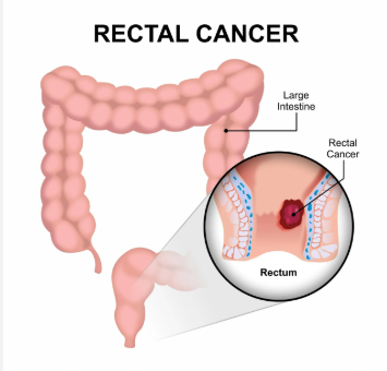
Whether your situation needs surgery alone, surgery with chemotherapy or something more complex involving multiple teams depends on your staging, your pathology and a multidisciplinary team that looks at your case as a whole rather than fitting it into a standard pathway.Rectal cancer treatment at specialist centres covers the full colorectal spectrum where every surgical plan gets built around what the specific case actually needs.
Why Choose Dr. Sandeep Nayak for Cancer Treatment?
Dr. Sandeep Nayak has been treating colon cancer surgically for over 24 years and doing D3 resection laparoscopically and robotically since before most Indian centres had made up their minds about minimally invasive colorectal surgery. MIND and RIA-MIND came out of operating in the precise anatomical planes that proper colon cancer surgery demands hundreds of times over rather than from a textbook. He chairs Oncology Services across Karnataka and sees patients at MACS Clinic in Bangalore. Dr. Nayak will look at your staging, your pathology and your specific case and build a plan from what’s actually there rather than from what the last similar patient needed.
Frequently Asked Questions
What is colon cancer? Cancer growing in the cells lining the large intestine, usually from polyps, with outcomes that depend heavily on the stage it’s caught at and the quality of treatment it gets.
What are the symptoms of colon cancer? Blood in stool, bowel habit that’s changed and stayed changed, unexplained weight loss, recurring abdominal pain and that feeling of never fully emptying the bowel.
How is colon cancer treated? Surgery with clear margins and lymph node removal as the foundation, chemotherapy when pathology shows it’s needed and minimally invasive approaches at specialist centres as standard.
Is colon cancer curable? Early stage colon cancer caught before it’s moved to nodes or distant sites is very often curable with surgery and proper follow-up at a specialist surgical oncology centre.
Robotic cancer surgery takes anywhere from two hours to eight or more depending on which cancer you’re having removed, how complex your anatomy is, whether lymph node dissection is part of what needs doing and whether previous surgery has left adhesions that need working through before anyone gets near the tumour, so anyone quoting you a single number without knowing your specific case is giving you an average that may have very little to do with how long you’ll actually be on the table.
According to Prof. Dr. Sandeep Nayak,Surgical Oncologist in India, “Operating time in robotic cancer surgery depends heavily on the specific procedure and the patient’s anatomy and quoting a single number without knowing the case doesn’t mean much.”
How Long Does Each Type of Robotic Cancer Surgery Take?
These are the approximate operating times for the most common robotic cancer procedures:
Thyroid cancer: RABIT and similar robotic thyroid approaches take two to four hours because the instruments travel through a tunnel from the armpit to the neck rather than going straight in, which adds setup and dissection time conventional open thyroid surgery simply doesn’t have.
Rectal cancer: Robotic low anterior resection and inter-sphincteric resection typically run three to five hours because the narrow pelvis, the sphincter preservation work and the precise dissection around nerves all take time that open surgery in a wider field doesn’t need to account for the same way.
Prostate cancer: Robotic prostatectomy runs two to four hours at experienced centres and the extra time compared to some open approaches goes into the nerve sparing work that makes the functional difference patients actually care about after surgery.
Colorectal cancer: Robotic hemicolectomy with D3 lymph node dissection takes two to four hours depending on tumour location, patient build and whether the vessel work during mesocolic dissection is straightforward or more involved than imaging suggested it would be.
High volume centres doing robotic cancer surgery regularly tend to run shorter operating times than lower volume centres and that’s not because they cut corners, it’s because familiarity with setup, anatomy and procedure steps means less time figuring things out mid-operation.Robotic cancer surgery at a specialist centre with a dedicated robotic team means the efficiency built from doing this repeatedly rather than occasionally is something you actually benefit from on the table.
What Factors Make Robotic Cancer Surgery Take Longer?
These are the things that push your operating time beyond the typical range for any robotic procedure:
Old scars inside: Adhesions from previous abdominal or pelvic surgery turn what should be clear tissue planes into a slow careful dissection and what normally takes an hour can easily take two if the adhesions are dense enough that rushing them creates a bleeding risk.
Tumour position: A tumour sitting right against a major vessel or in a location that limits how the instruments can approach it takes longer to remove safely regardless of how experienced the surgeon is because the physics of the situation don’t change with experience.
Lymph node work: Extended lymphadenectomy like D3 resection or central neck dissection adds real time to any robotic procedure because clearing node basins at vessel origins is systematic careful work that can’t be done faster without making it less thorough.
Your build and anatomy: Obesity, a narrow pelvis, a short thick neck or other anatomical factors that reduce the working space for robotic instruments consistently push operating time up because the surgeon is working in a smaller environment than the time estimates assume.
What your specific procedure is likely to take is something your surgical team can estimate much more accurately once they’ve looked at your imaging and understood your anatomy rather than giving you a generic number from a website.Laparoscopic cancer surgery at specialist centres covers the full minimally invasive range where operating time and case complexity get individually assessed before anything gets booked.
Why Choose Dr. Sandeep Nayak for Cancer Treatment?
Dr. Sandeep Nayak has been doing robotic cancer surgery for over 15 years and the efficiency of his team at MACS Clinic in Bangalore comes from doing this at real volume rather than occasionally, which matters because time under anaesthesia is itself a risk factor especially for older patients or those carrying other health conditions going into surgery. He chairs Oncology Services across Karnataka. Dr. Nayak will tell you realistically how long your procedure is likely to run, what drives that estimate for your specific case and what it means for your recovery rather than quoting a reassuring number that has nothing to do with what’s actually on your scan.
Frequently Asked Questions
How long does robotic cancer surgery take? Two to eight hours typically depending on cancer type, procedure complexity, lymph node dissection and patient anatomy.
Why does robotic surgery sometimes take longer than open surgery? Setup time, tunnel approaches for certain procedures and the precision dissection that makes robotic surgery worth doing all add time open incisions don’t require.
Does previous surgery affect how long robotic cancer surgery takes? Yes, internal adhesions from prior operations slow dissection significantly and can add one to two hours beyond the typical range for the procedure.
Does time on the table affect recovery after robotic cancer surgery? Yes, longer anaesthesia and operating time increases risk and recovery complexity particularly for older patients or those with existing health conditions going in.
Dr. Sandeep Nayak, a board-certified surgical oncologist (M.ch ., D.N.B.), specializes in minimally invasive cancer surgery, including robotic and laparoscopic techniques.